. 2008 Nov 15;181(10):7214-20.
doi: 10.4049/jimmunol.181.10.7214.

IL-10 and TGF-beta redundantly protect against severe liver injury and mortality during acute schistosomiasis

Affiliations

IL-10 and TGF-beta redundantly protect against severe liver injury and mortality during acute schistosomiasis

De'Broski R Herbert et al. J Immunol. .

Abstract

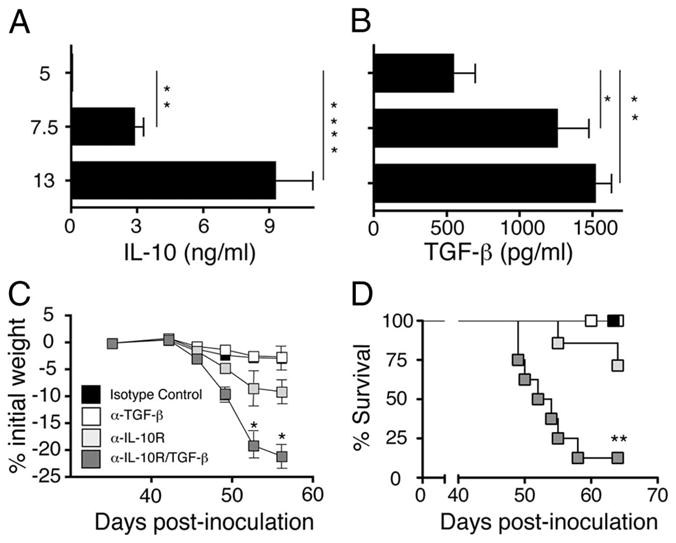
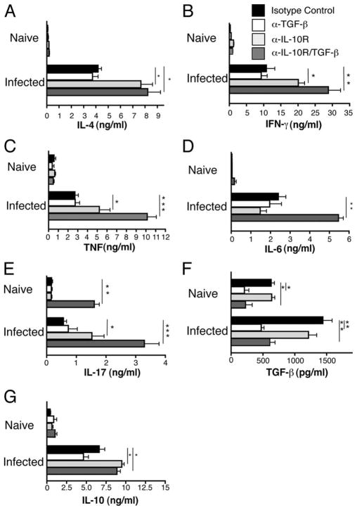
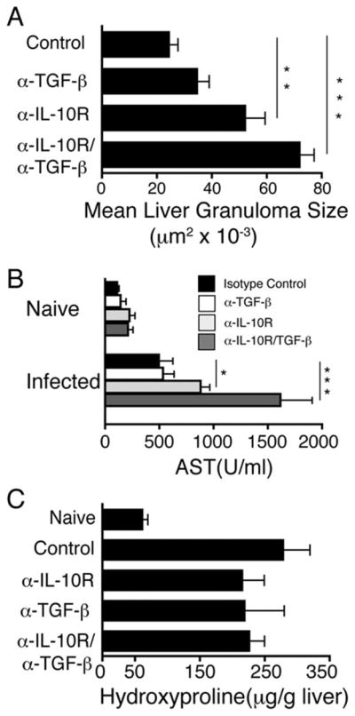
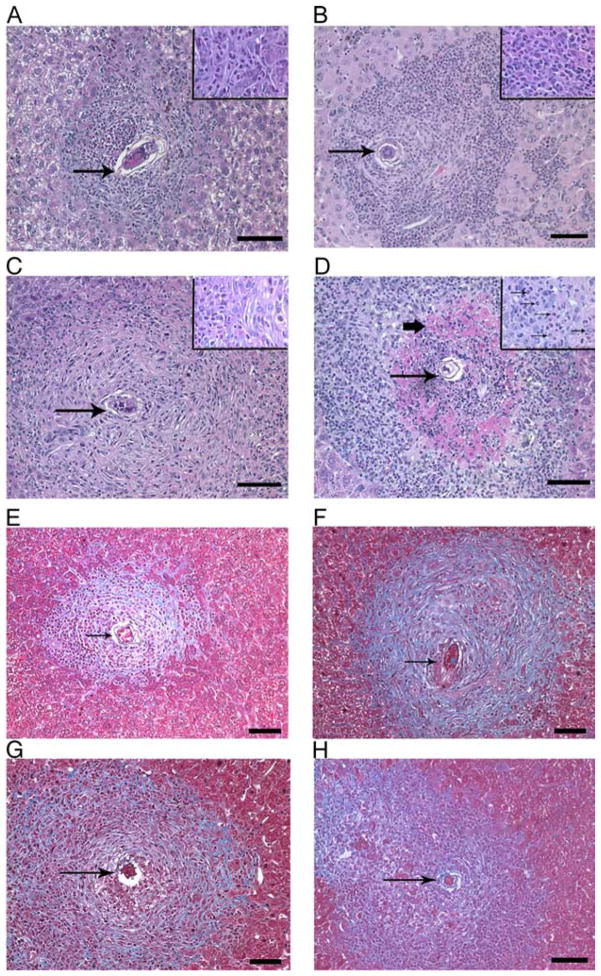
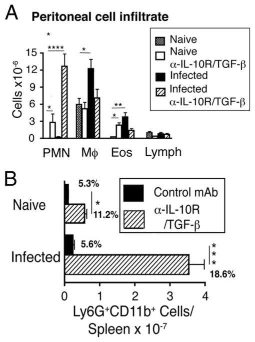
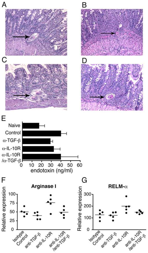
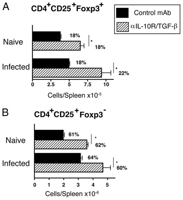
The cytokines IL-10 and TGF-beta regulate immunity and inflammation. IL-10 is known to suppress the extent of hepatic damage caused by parasite ova during natural infection with Schistosoma mansoni, but the role of TGF-beta is less clear. Cytokine blockade studies in mice revealed that anti-IL-10R mAb treatment during acute infection modestly increased cytokine production and liver damage, whereas selective anti-TGF-beta mAb treatment had marginal effects. In contrast, mice administered both mAbs developed severe hepatic inflammation, with enlarged, necrotic liver granulomas, cachexia, and >80% mortality by 8 wk postinfection, despite increased numbers of CD4(+)CD25(+)Foxp3(+) T regulatory cells. Blocking both IL-10 and TGF-beta at the onset of egg production also significantly increased IL-4, IL-6, TNF, IFN-gamma, and IL-17 production and markedly increased hepatic, peritoneal, and splenic neutrophilia. In contrast, coadministration of anti-IL-10R and TGF-beta mAbs had little effect upon parasite ova-induced intestinal pathology or development of alternatively activated macrophages, which are required to suppress intestinal pathology. This suggests that inflammation is controlled during acute S. mansoni infection by two distinct, organ-specific mechanisms: TGF-beta and IL-10 redundantly suppress hepatic inflammation while intestinal inflammation is regulated by alternatively activated macrophages.


Conflict of interest statement

Disclosures

The authors have no financial conflict of interest.

Figures

FIGURE 1
FIGURE 1
IL-10 and TGF-β redundantly protect against lethal schistosomiasis. Serum levels of IL-10 (A) and TGF-β (B) determined by IVCCA and ELISA, respectively, at the indicated time points in mice infected with 60–70 S. mansoni cercariae. Means ± SE of 8–10 mice/group. Experiment performed twice. Weight change (C) and survival kinetics (D) of BALB/c mice infected with 60–70 S. mansoni cercariae were injected i.p on days 38, 42, and 46 with 1 mg of anti-TGF-β mAb (clone 1D11), 1 mg of anti-IL-10 receptor mAb (1B1.3), 1 mg each of both mAbs, or 2 mg of an isotype control mAb (clone J1.2). Data shown as means ± SE of eight mice/group. Experiment performed four times (*, p < 0.05; **, p < 0.01; and ***, p < 0.0001 compared with isotype control treated group). Significance determined by ANOVA.
FIGURE 2
FIGURE 2
Blocking IL-10 and TGF-β causes increased levels of cytokines associated with TH1, TH2, and TH17 polarization. Production of (A) IL-4, (B) IFN-γ, (C) TNF, (D) IL-6 (G), IL-10 (measured by IVCCA), (E) IL-17, and (F) TGF-β1 (measured by ELISA) in sera from naive or S. mansoni-inoculated BALB/c mice on day 52 postinfection. Animals were injected i.p. on days 38, 42, and 46 with 1 mg of anti-TGF-β (clone 1D11), 1 mg of anti-IL-10 receptor (1B1.3), 1 mg of both anti-TGF-β/anti-IL-10R, or 2 mg of isotype control (clone J1.2). Means ± SE of six to eight mice/group. Experiment performed three times (*, p < 0.05; **, p < 0.01, and ***, p < 0.0001 compared with isotype control treated group).
FIGURE 3
FIGURE 3
Blocking both IL-10 and TGF-β exacerbates hepatocellular damage and granulomatous pathology, but does not affect fibrosis. A, Liver granuloma cross-sectional area was determined 52 days postinoculation. 300 granulomas were measured per group. B, Serum levels of AST in naive or S. mansoni infected mice 52 days postinfection. C, Liver hydroxyproline levels 52 days postinfection. Means ± SE of six to eight mice/group. Experiments performed three times with similar results (*, p < 0.05; **, p < 0.01; and ***, p < 0.001 compared with isotype control-treated group).
FIGURE 4
FIGURE 4
Blocking both IL-10 and TGF-β causes hepatic necrosis. Liver sections from S. mansoni-inoculated mice on day 52 postinfection following treatment with isotype control (A and E), anti-TGF-β (B and F), anti-IL-10R (C and G), or anti-TGF-β/anti-IL-10R (D and H) Ab. Thin arrows point to parasite ova. Bold arrow points to area of necrosis. Representative photos are shown. A–D, H&E-stained sections. ×100 magnification (large panel) 600× (inset) Scale bar 100 μm. E–H, Masson’s trichrome-stained sections at a ×200 magnification. Staining for collagen (blue) and hepatocytes (reddish/purple). Scale bar 50 μm.
FIGURE 5
FIGURE 5
Blocking IL-10 and TGF-β during acute schistosomiasis causes profound neutrophilia in peritoneal cavity and spleen. A, Inflammatory cells from peritoneal lavage from naive or S. mansoni-inoculated mice (day 52) treated with 1 mg each of anti-IL-10R/TGF-β mAbs or left untreated. B, Spleen cells from naive or S. mansoni-inoculated mice (day 52) were stained for Ly6G/C (GR-1+) and Mac-1 (CD11b+) following injection with 1 mg of anti-IL-10R/TGF-β or control mAbs. Means ± SE of total numbers per spleen from four mice/group. Percent gated population shown on graph. Experiments performed twice with similar results (*, p < 0.05; **, p < 0.01; ***, p < 0.001; and ****, p < 0.0001 compared with isotype control treated group).
FIGURE 6
FIGURE 6
Blocking IL-10 and TGF-β does not affect intestinal pathology, alternative macrophage activation, or mucosal integrity. H&E staining of ileal tissue sections from S. mansoni infected mice on d 52 postinfection following treatment with isotype control (A), anti-TGF-β (B), anti-IL-10R (C), or anti-TGF-β/anti-IL-10R (D) mAb. Magnification 200×. Arrows point to parasite ova. Measurement of serum endotoxin levels on day 52 postinfection (E). Real-time PCR quantification of gut mRNA transcripts for (F) Arginase I and (G) RELM-α. Means ± SE of five mice/group. Experiment performed twice.
FIGURE 7
FIGURE 7
Blocking IL-10 and TGF-β during acute schistosomiasis infection increases Treg number. Spleen cells from naive or S. mansoni-inoculated mice (day 52) that received 2 mg of GL113 or were treated with 1 mg each of anti-IL-10R/TGF-β mAbs were stained with fluorochrome-labeled mAbs for CD4 (RM4-5), CD25 (PC-61), and Foxp3 (FJK-16s) and analyzed by flow cytometry for number of CD4+CD25+Foxp3+ cells. Percent of gated CD4+ cell population shown on graph. Means ± SE of four mice/group. Experiments were performed twice (A and B) with similar results (*, p < 0.05; **, p < 0.01; and ***, p < 0.001 compared with isotype control-treated group).

References

    1. Wan YY, Flavell RA. “Yin-Yang” functions of transforming growth factor-β and T regulatory cells in immune regulation. Immunol Rev. 2007;220:199–213. - PMC - PubMed
    1. O’Garra A, Vieira PL, Vieira P, Goldfeld AE. IL-10-producing and naturally occurring CD4+ Tregs: limiting collateral damage. J Clin Invest. 2004;114:1372–1378. - PMC - PubMed
    1. Dunne DW, Pearce EJ. Immunology of hepatosplenic schistosomiasis mansoni: a human perspective. Microbes Infect. 1999;1:553–560. - PubMed
    1. Abath FG, Morais CN, Montenegro CE, Wynn TA, Montenegro SM. Immunopathogenic mechanisms in schistosomiasis: what can be learnt from human studies? Trends Parasitol. 2006;22:85–91. - PubMed
    1. Stadecker MJ. The shrinking schistosomal egg granuloma: how accessory cells control T cell-mediated pathology. Exp Parasitol. 1994;79:198–201. - PubMed

Publication types

MeSH terms

Substances