. 2008 Nov 3;183(3):377-84.
doi: 10.1083/jcb.200808137.

Deletion of IFT20 in the mouse kidney causes misorientation of the mitotic spindle and cystic kidney disease

Affiliations

Deletion of IFT20 in the mouse kidney causes misorientation of the mitotic spindle and cystic kidney disease

Julie A Jonassen et al. J Cell Biol. .

Abstract

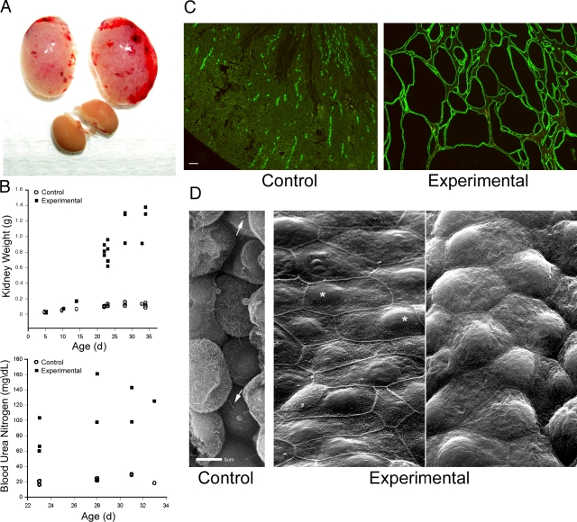
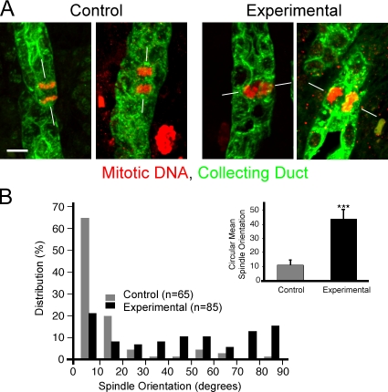
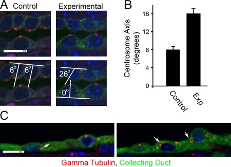
Primary cilia project from the surface of most vertebrate cells and are thought to be sensory organelles. Defects in primary cilia lead to cystic kidney disease, although the ciliary mechanisms that promote and maintain normal renal function remain incompletely understood. In this work, we generated a floxed allele of the ciliary assembly gene Ift20. Deleting this gene specifically in kidney collecting duct cells prevents cilia formation and promotes rapid postnatal cystic expansion of the kidney. Dividing collecting duct cells in early stages of cyst formation fail to properly orient their mitotic spindles along the tubule, whereas nondividing cells improperly position their centrosomes. At later stages, cells lacking cilia have increased canonical Wnt signaling and increased rates of proliferation. Thus, IFT20 functions to couple extracellular events to cell proliferation and differentiation.


Figures

Figure 1.
Figure 1.
Deletion of Ift20 in mouse kidney. (A) Gross morphology of experimental kidneys (top pair) and control kidneys at p23. (B) Mean weight of kidneys and blood urea nitrogen levels of control and experimental animals. Each symbol represents one animal. The symbols at 5 and 10 d were slightly offset. (C) Adult (p23) kidneys stained with DBA (green). (D) Scanning EM images of control and experimental kidneys. Arrows indicate cilia and asterisks indicate nuclei. Bars: (C) 100 μm; (D) 5 μm.
Figure 2.
Figure 2.
Relationship between cilia loss and cystic expansion. Kidneys from control and experimental animals were stained for cilia (611β1, green), collecting ducts (aquaporin-2, red), and nuclei (DAPI, blue) at e18, which is the day before birth, and p5, p10, and p23. Arrows indicate collecting ducts and arrowheads indicate ciliated nephrons near collecting ducts; cysts and dilated ducts are indicated by a “C.” Images are maximum projections of 16 confocal z images taken 0.5 μm apart. Bar, 10 μm.
Figure 3.
Figure 3.
Deletion of Ift20 leads to mitotic spindle misorientation in p5 collecting duct cells. (A) In normal kidney tubules, mitotic spindles (phospho-histone H3, red) typically orient their axes (white lines) parallel to the long axis of the collecting duct (DBA, green; left panels in A, gray bars in B). The absence of IFT20 disrupts this arrangement, producing a more random orientation of mitotic spindles (right panels in A, black bars in B). Images are maximum projections of 16 confocal z images taken 0.5 μm apart. Bar, 5 μm. (B) Mitotic spindle orientation quantitation. Mitotic collecting duct cells were photographed, and the angle between the long axis of the tubule and the spindle was measured; angles were grouped into 10° bins. Bars in the inset show circular mean mitotic spindle orientation and the 95% confidence interval about the mean. ***, P < 0.001 (Kolmogorov-Smirnov test).
Figure 4.
Figure 4.
Centrosome defects. (A) In control p5 kidneys, centrosomes (γ-tubulin, red) are typically found at the center of the apical surface of collecting duct cells, whereas the position in experimental animals is more variable. The position was quantified by measuring the angle between a line along the basal surface of the cell and a line drawn between the centrosome and the center of the cell at the basal surface. The cell center was defined by the center of the nucleus. The bottom two panels illustrate this measurement. Images are maximum projections of four confocal z images taken 0.5 μm apart. (B) Quantification of the centrosome axis. n = 100. Error bars indicate SEM. (C) Two examples of the unusual localization of centrosomes (arrows) that can be found in highly cystic kidneys. Bars, 10 μm.
Figure 5.
Figure 5.
Deletion of Ift20 alters canonical Wnt signaling. (A) β-catenin staining of control (top rows) and experimental (bottom rows) kidneys at p10 and p33. Arrows indicate collecting ducts in controls and dilated or cystic ducts in experimental mice. Images are maximum projections of five confocal z images taken 0.5 μm apart (2 μm total). Control and experimental images at each of the time points were taken under identical conditions and manipulated uniformly, but the conditions were not the same at p10 and p33. Bar, 10 μm. (B) Western blot analysis of β-catenin. Control (Con) and experimental (Exp) kidneys were separated into cytosol and nuclear fractions and analyzed by Western blots with antibodies to β-catenin and dephosphorylated (active) β-catenin. RelB and GAPDH were used as loading controls. “1” and “2” represent different animals. (C) qPCR analysis of nine genes in experimental (closed bars) and control (open bars) kidneys at selected postnatal times. Gene expression data are normalized to GAPDH expression. Bars indicate mean ± SEM of 5–11 mice in each treatment and age group, plotted on logarithmic scales. *, P < 0.05; **, P < 0.01 (unpaired t tests).

References

    1. Chuang, Y.Y., A. Valster, S.J. Coniglio, J.M. Backer, and M. Symons. 2007. The atypical Rho family GTPase Wrch-1 regulates focal adhesion formation and cell migration. J. Cell Sci. 120:1927–1934. - PubMed
    1. Corbit, K.C., A.E. Shyer, W.E. Dowdle, J. Gaulden, V. Singla, and J.F. Reiter. 2008. Kif3a constrains beta-catenin-dependent Wnt signalling through dual ciliary and non-ciliary mechanisms. Nat. Cell Biol. 10:70–76. - PubMed
    1. Daniel, W.W. 1990. Applied Nonparametric Statistics. Second Edition. Pacific Grove, CA: Duxbury Thomson Learning. 635 pp.
    1. Davenport, J.R., A.J. Watts, V.C. Roper, M.J. Croyle, T. van Groen, J.M. Wyss, T.R. Nagy, R.A. Kesterson, and B.K. Yoder. 2007. Disruption of intraflagellar transport in adult mice leads to obesity and slow-onset cystic kidney disease. Curr. Biol. 17:1586–1594. - PMC - PubMed
    1. Farley, F.W., P. Soriano, L.S. Steffen, and S.M. Dymecki. 2000. Widespread recombinase expression using FLPeR (flipper) mice. Genesis. 28:106–110. - PubMed

Publication types

MeSH terms