. 2008 Nov 1;22(21):2941-52.
doi: 10.1101/gad.1709008.

PPARgamma and C/EBP factors orchestrate adipocyte biology via adjacent binding on a genome-wide scale

Affiliations

PPARgamma and C/EBP factors orchestrate adipocyte biology via adjacent binding on a genome-wide scale

Martina I Lefterova et al. Genes Dev. .

Abstract

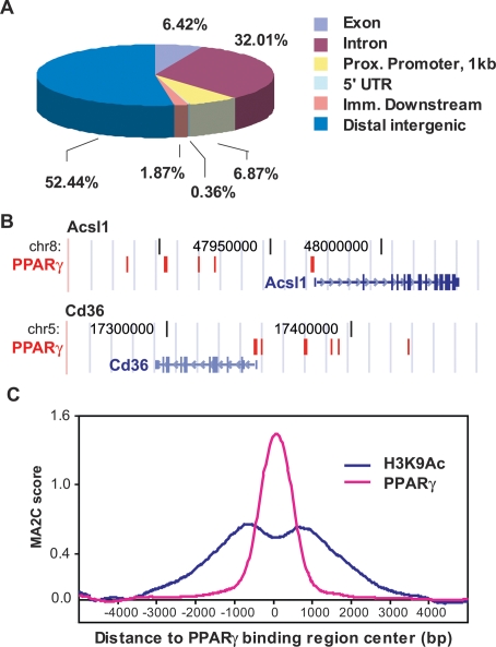
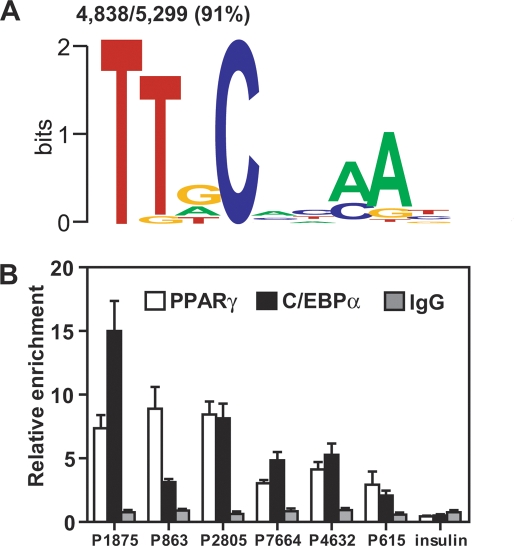
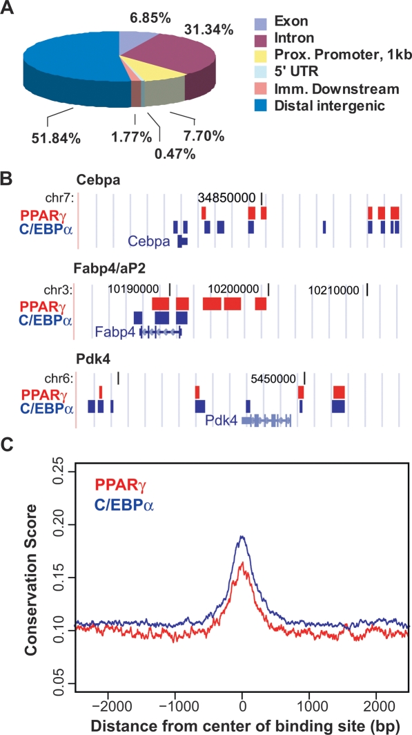
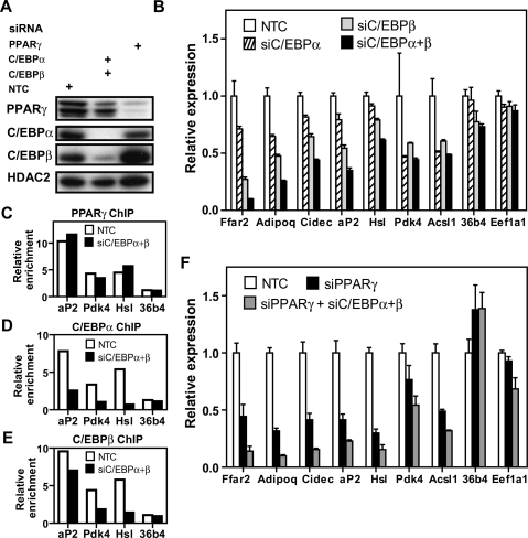
Peroxisome proliferator-activated receptor gamma(PPARgamma), a nuclear receptor and the target of anti-diabetic thiazolinedione drugs, is known as the master regulator of adipocyte biology. Although it regulates hundreds of adipocyte genes, PPARgamma binding to endogenous genes has rarely been demonstrated. Here, utilizing chromatin immunoprecipitation (ChIP) coupled with whole genome tiling arrays, we identified 5299 genomic regions of PPARgamma binding in mouse 3T3-L1 adipocytes. The consensus PPARgamma/RXRalpha "DR-1"-binding motif was found at most of the sites, and ChIP for RXRalpha showed colocalization at nearly all locations tested. Bioinformatics analysis also revealed CCAAT/enhancer-binding protein (C/EBP)-binding motifs in the vicinity of most PPARgamma-binding sites, and genome-wide analysis of C/EBPalpha binding demonstrated that it localized to 3350 of the locations bound by PPARgamma. Importantly, most genes induced in adipogenesis were bound by both PPARgamma and C/EBPalpha, while very few were PPARgamma-specific. C/EBPbeta also plays a role at many of these genes, such that both C/EBPalpha and beta are required along with PPARgamma for robust adipocyte-specific gene expression. Thus, PPARgamma and C/EBP factors cooperatively orchestrate adipocyte biology by adjacent binding on an unanticipated scale.


Figures

Figure 1.
Figure 1.
Location analysis of PPARγ-binding sites. (A) PPARγ-binding regions were mapped relative to their nearest RefSeq genes using CEAS (Ji et al. 2006). Proximal (prox.) promoter was defined as ≤1 kb upstream from the TSS. Immediate (imm.) downstream was defined as ≤1 kb downstream from the 3′ end of the gene. Distal intergenic refers to all locations outside the boundaries of a gene and the 1 kb flanking the gene on either end. (UTR) Untranslated region. (B) PPARγ-binding regions are frequently clustered around target genes. Two PPARγ target genes, Cd36 and Acsl1, are shown in their native chromosomal locations according to the February 2006 Mouse Genome Assembly (mm8) in the UCSC Genome Browser (http://genome.ucsc.edu). Red blocks represent regions of enriched PPARγ-binding signal. Vertical lines within the genes represent exons, horizontal lines represent introns, and arrowheads represent the direction of transcription. (C) Enrichment of acetylation at Lys 9 of histone 3 (H3K9Ac) in the regions of PPARγ binding. Shown are the average ChIP–chip profiles for PPARγ and H3K9Ac across 740 PPARγ-binding regions located >10 kb from a TSS. MA2C score refers to the enrichment at each location along the 10 kb distance that was tiled on the custom array for each region.
Figure 2.
Figure 2.
RXRα heterodimerization and DR1 enrichment at novel PPARγ-binding sites. (A) Overlap in binding between PPARγ and RXRα across 1431 PPARγ-binding regions identified previously in the genome-wide search and interrogated in the custom “PPARγ-binding site” arrays. Shown in parentheses is the number of enriched regions for each antibody. (B,C) Enriched motif analysis of the PPARγ sites using TRANSFAC and JASPAR PWMs. (B) A DR1-like element was found in 75% of the sites. (C) Ninety-six percent of the novel PPARγ-binding regions contain at least the half site of the consensus PPARγ response element.
Figure 3.
Figure 3.
C/EBP response elements are found at the vast majority of PPARγ-binding regions. (A) Enrichment of C/EBP motifs. The PPARγ-binding locations were mined as in Figure 2, B and C. Shown is the logo of one C/EBP PWM among several that were enriched. (B) ChIP-QPCR analysis for C/EBPα and PPARγ at several novel PPARγ-binding regions that were computationally predicted to contain C/EBP response elements (see Supplemental Tables 1 and 5 for identification and location of the PPARγ-binding sites). An area of the insulin gene served as negative control for PPARγ and C/EBPα binding. Data are normalized to a site in the Arbp/36b4 gene and presented as mean ± SE, n = 3.
Figure 4.
Figure 4.
Location analysis of C/EBPα binding. (A) Mapping of C/EBPα-binding regions on genome-wide scale relative to RefSeq mouse genes. The analysis was performed as in Figure 1A. (B) C/EBPα and PPARγ binding in relation to three target genes, Cebpa, Fabp4/aP2, and Pdk4. The genes are shown as in Figure 1B, in the native chromosomal locations according to the mm8 Assembly in the UCSC Genome Browser (http://genome.ucsc.edu). Blue blocks represent regions of C/EBPα enriched ChIP signal, while red blocks represent PPARγ enrichment. (C) Average plot for conservation of all PPARγ- and C/EBPα-binding regions among higher eukaryotes.
Figure 5.
Figure 5.
Extent of PPARγ and C/EBPα binding overlap and its association with gene expression during adipocyte differentiation. (A) Overlap between the binding of PPARγ and C/EBPα on genome-wide scale. Shown are the numbers of regions found to be shared by the two factors—i.e., having at least 1 bp in common—or unique to each factor. (B) Summary of gene ontology (GO) categories of the nearest genes to regions with overlapping PPARγ and C/EBPα binding. In this analysis, only binding regions whose nearest gene was within 50 kb were considered. (C) Association between factor binding and genes induced in adipogenesis. Shown are the percent genes up-regulated more than threefold and containing binding sites within 50 kb of the gene start site for both factors (Both), PPARγ alone, C/EBPα alone, or neither factor (Neither). (D) The association between genes down-regulated in adipocyte differentiation and PPARγ and C/EBPα binding was analyzed as in C.
Figure 6.
Figure 6.
Effects of C/EBP depletion on expression of genes on which PPARγ and the C/EBPs colocalize. (A) Immunoblot analysis demonstrating the efficiency of siRNA-mediated knockdown of C/EBPα, C/EBPβ, PPARγ, or nontarget contol (NTC). HDAC2 represents a loading control. (B) QPCR analysis of gene expression following 24 h of siRNA-mediated knockdown. All of the genes shown were found to have binding sites for PPARγ and C/EBPα, except eukaryotic translation elongation factor 1 α 1 (Eef1α1) and 36b4, which were used as controls. Data were normalized to the housekeeping gene Pabpc1, and are presented as mean ± SE, n = 3. (C–E) ChIP-QPCR analysis of factor binding at several target sites following 24 h of C/EBPα and β or NTC knockdown. Data are normalized to a nontarget genomic site and IgG enrichment. Shown is a representative ChIP-QPCR experiment. (C) PPARγ enrichment. (D) C/EBPα enrichment. (E) C/EBPβ enrichment. (F) QPCR analysis of gene expression following 24 h of siRNA-mediated knockdown of PPARγ alone and together with C/EBPα and C/EBPβ. Analysis was performed as in B above.

References

    1. Anderson P.D., Mehta N.N., Wolfe M.L., Hinkle C.C., Pruscino L., Comiskey L.L., Tabita-Martinez J., Sellers K.F., Rickels M.R., Ahima R.S., et al. Innate immunity modulates adipokines in humans. J. Clin. Endocrinol. Metab. 2007;92:2272–2279. - PubMed
    1. Bolton E.C., So A.Y., Chaivorapol C., Haqq C.M., Li H., Yamamoto K.R. Cell- and gene-specific regulation of primary target genes by the androgen receptor. Genes & Dev. 2007;21:2005–2017. - PMC - PubMed
    1. Cao Z., Umek R.M., McKnight S.L. Regulated expression of three C/EBP isoforms during adipose conversion of 3T3-L1 cells. Genes & Dev. 1991;5:1538–1552. - PubMed
    1. Carroll J.S., Liu X.S., Brodsky A.S., Li W., Meyer C.A., Szary A.J., Eeckhoute J., Shao W., Hestermann E.V., Geistlinger T.R., et al. Chromosome-wide mapping of estrogen receptor binding reveals long-range regulation requiring the forkhead protein FoxA1. Cell. 2005;122:33–43. - PubMed
    1. Carroll J.S., Meyer C.A., Song J., Li W., Geistlinger T.R., Eeckhoute J., Brodsky A.S., Keeton E.K., Fertuck K.C., Hall G.F., et al. Genome-wide analysis of estrogen receptor binding sites. Nat. Genet. 2006;38:1289–1297. - PubMed

Publication types

Substances