

(18)F and (18)FDG PET imaging of osteosarcoma to non-invasively monitor in situ changes in cellular proliferation and bone differentiation upon MYC inactivation
- PMID: 18981708
- PMCID: PMC4158945
- DOI: 10.4161/cbt.7.12.6947
(18)F and (18)FDG PET imaging of osteosarcoma to non-invasively monitor in situ changes in cellular proliferation and bone differentiation upon MYC inactivation
Abstract
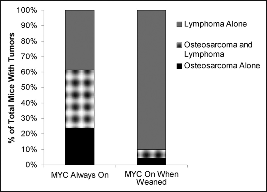
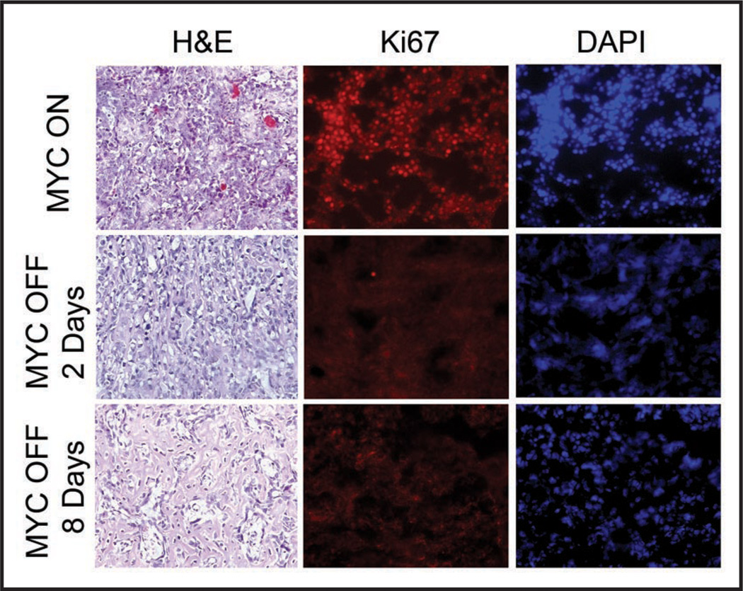
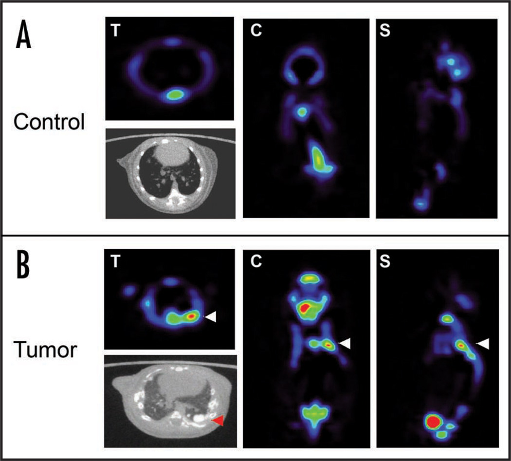
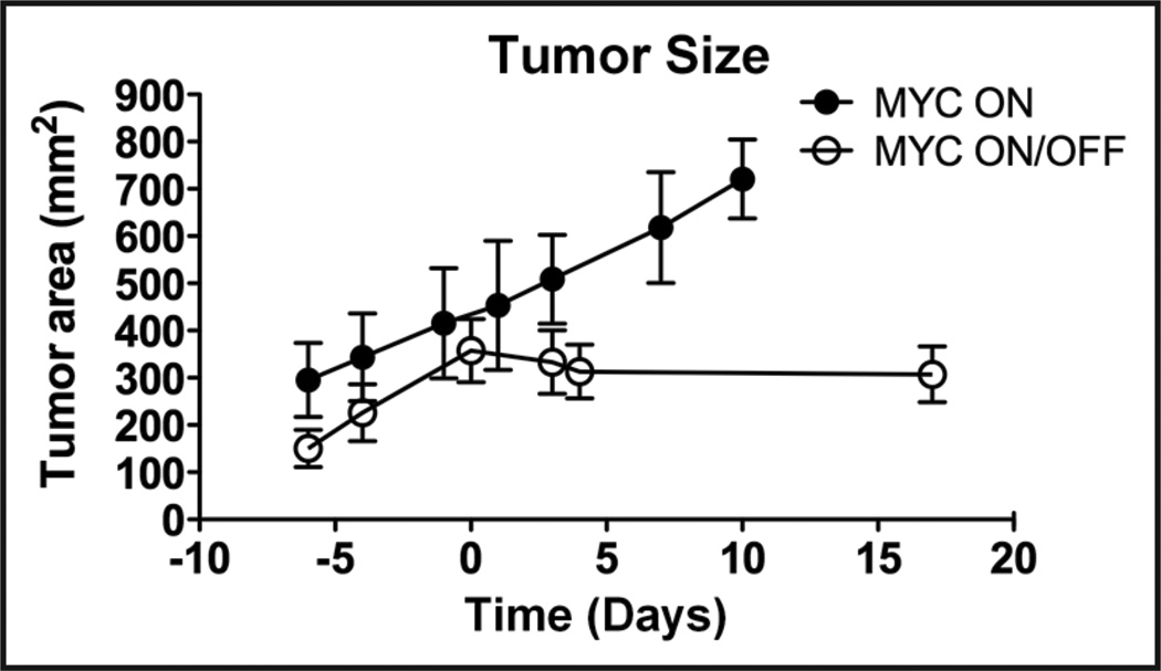
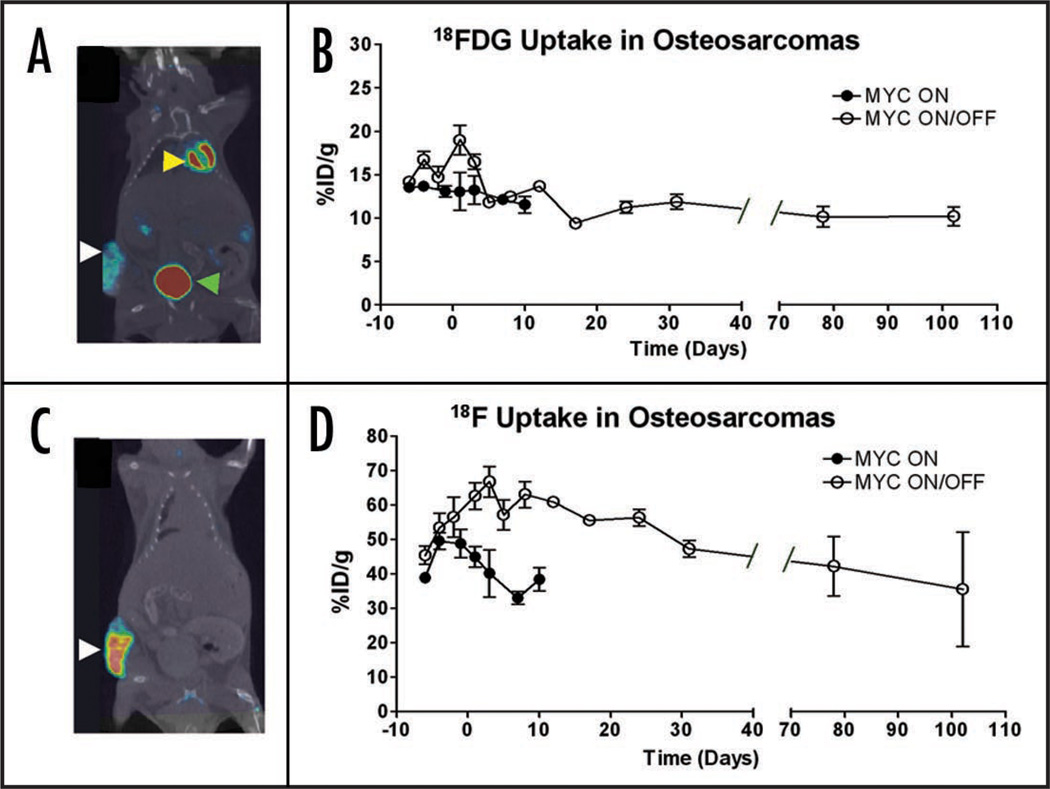
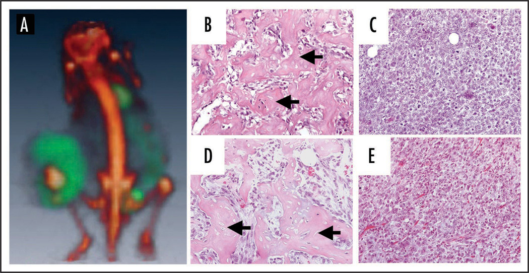
Osteosarcoma is one of the most common pediatric cancers. Accurate imaging of osteosarcoma is important for proper clinical staging of the disease and monitoring of the tumor's response to therapy. The MYC oncogene has been commonly implicated in the pathogenesis of human osteosarcoma. Previously, we have described a conditional transgenic mouse model of MYC-induced osteosarcoma. These tumors are highly invasive and are frequently associated with pulmonary metastases. In our model, upon MYC inactivation osteosarcomas lose their neoplastic properties, undergo proliferative arrest and differentiate into mature bone. We reasoned that we could use our model system to develop noninvasive imaging modalities to interrogate the consequences of MYC inactivation on tumor cell biology in situ. We performed positron emission tomography (PET) combining the use of both (18)F-fluorodeoxyglucose ((18)FDG) and (18)F-flouride ((18)F) to detect metabolic activity and bone mineralization/remodeling. We found that upon MYC inactivation, tumors exhibited a slight reduction in uptake of (18)FDG and a significant increase in the uptake of (18)F along with associated histological changes. Thus, these cells have apparently lost their neoplastic properties based upon both examination of their histology and biologic activity. However, these tumors continue to accumulate (18)FDG at levels significantly elevated compared to normal bone. Therefore, PET can be used to distinguish normal bone cells from tumors that have undergone differentiation upon oncogene inactivation. In addition, we found that (18)F is a highly sensitive tracer for detection of pulmonary metastasis. Collectively, we conclude that combined modality PET/CT imaging incorporating both (18)FDG and (18)F is a highly sensitive means to non-invasively measure osteosarcoma growth and the therapeutic response, as well as to detect tumor cells that have undergone differentiation upon oncogene inactivation.
Figures
References
-
- Bruland OS, Pihl A. On the current management of osteosarcoma. A critical evaluation and a proposal for a modified treatment strategy. Eur J Cancer. 1997;33:1725–1731. - PubMed
-
- Dimitrakopoulou-Strauss A, Hohenberger P, Strobel P, Marx A, Strauss LG. A recent application of fluoro-18-deoxyglucose positron emission tomography, treatment monitoring with a mammalian target of rapamycin inhibitor: an example of a patient with a desmoplastic small round cell tumor. Hell J Nucl Med. 2007;10:77–79. - PubMed
-
- Kumar R, Chauhan A, Kesav Vellimana A, Chawla M. Role of PET/PET-CT in the management of sarcomas. Expert Rev Anticancer Ther. 2006;6:1241–1250. - PubMed
Publication types
MeSH terms
Substances
Grants and funding
LinkOut - more resources
Full Text Sources
Medical