

MHC class II-expressing thymocytes suppress invariant NKT cell development
- PMID: 18982019
- PMCID: PMC2801351
- DOI: 10.1038/icb.2008.78
MHC class II-expressing thymocytes suppress invariant NKT cell development
Abstract
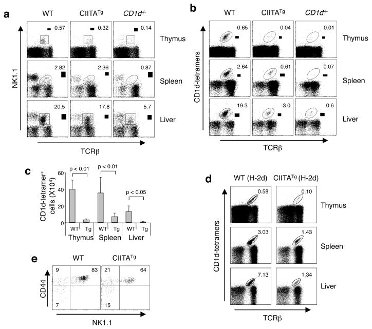
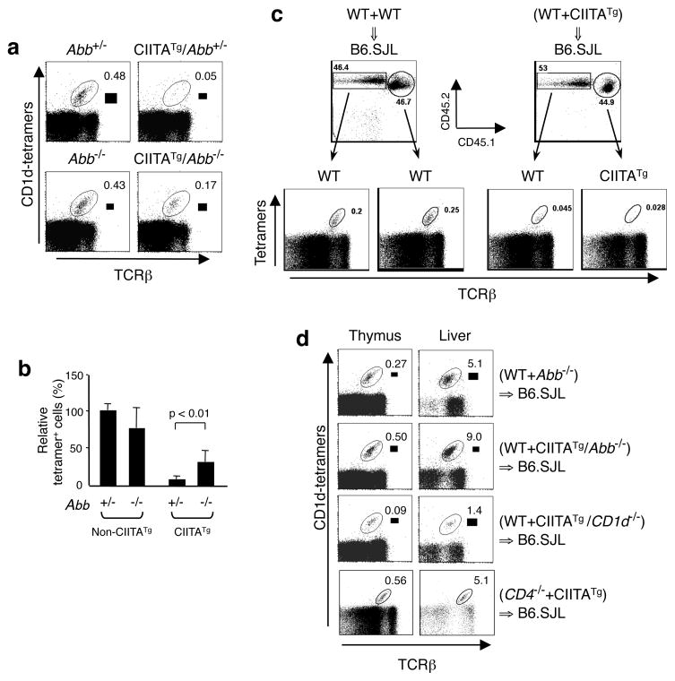
Natural killer T (NKT) cells are positively selected on cortical thymocytes expressing the non-classical major histocompatibility complex (MHC) class I CD1d molecules. However, it is less clear how NKT cells are negatively selected in the thymus. In this study, we investigated the role of MHC class II expression in NKT cell development. Transgenic mice expressing MHC class II on thymocytes and peripheral T cells had a marked reduction in invariant NKT (iNKT) cells. Reduced numbers of iNKT cells correlated with the absence of in vivo production of cytokines in response to the iNKT cell agonist alpha-galactosylceramide. Using mixed bone marrow chimeras, we found that MHC class II-expressing thymocytes suppressed the development of iNKT cells in trans in a CD4-dependent manner. Our observations have significant implications for human iNKT cell development as human thymocytes express MHC class II, which can lead to an inefficient selection of iNKT cells.
Figures
Similar articles
-
Classical MHC expression by DP thymocytes impairs the selection of non-classical MHC restricted innate-like T cells.Nat Commun. 2021 Apr 16;12(1):2308. doi: 10.1038/s41467-021-22589-z. Nat Commun. 2021. PMID: 33863906 Free PMC article.
-
Generation of PLZF+ CD4+ T cells via MHC class II-dependent thymocyte-thymocyte interaction is a physiological process in humans.J Exp Med. 2010 Jan 18;207(1):237-46. doi: 10.1084/jem.20091519. Epub 2009 Dec 28. J Exp Med. 2010. PMID: 20038602 Free PMC article.
-
Critical role for invariant chain in CD1d-mediated selection and maturation of Vα14-invariant NKT cells.Immunol Lett. 2011 Sep 30;139(1-2):33-41. doi: 10.1016/j.imlet.2011.04.012. Epub 2011 May 5. Immunol Lett. 2011. PMID: 21565221 Free PMC article.
-
New Genetically Manipulated Mice Provide Insights Into the Development and Physiological Functions of Invariant Natural Killer T Cells.Front Immunol. 2018 Jun 14;9:1294. doi: 10.3389/fimmu.2018.01294. eCollection 2018. Front Immunol. 2018. PMID: 29963043 Free PMC article. Review.
-
The ins and outs of type I iNKT cell development.Mol Immunol. 2019 Jan;105:116-130. doi: 10.1016/j.molimm.2018.09.023. Epub 2018 Nov 28. Mol Immunol. 2019. PMID: 30502719 Free PMC article. Review.
Cited by
-
Classical MHC expression by DP thymocytes impairs the selection of non-classical MHC restricted innate-like T cells.Nat Commun. 2021 Apr 16;12(1):2308. doi: 10.1038/s41467-021-22589-z. Nat Commun. 2021. PMID: 33863906 Free PMC article.
-
A cytokine-delivering polymer is effective in reducing tumor burden in a head and neck squamous cell carcinoma murine model.Otolaryngol Head Neck Surg. 2014 Sep;151(3):447-53. doi: 10.1177/0194599814533775. Epub 2014 May 13. Otolaryngol Head Neck Surg. 2014. PMID: 24825873 Free PMC article.
-
Generation of PLZF+ CD4+ T cells via MHC class II-dependent thymocyte-thymocyte interaction is a physiological process in humans.J Exp Med. 2010 Jan 18;207(1):237-46. doi: 10.1084/jem.20091519. Epub 2009 Dec 28. J Exp Med. 2010. PMID: 20038602 Free PMC article.
References
-
- Bendelac A, Savage PB, Teyton L. The biology of NKT cells. Annu Rev Immunol. 2007;25:297–336. - PubMed
-
- Kronenberg M. Toward an understanding of NKT cell biology: progress and paradoxes. Annu Rev Immunol. 2005;23:877–900. - PubMed
-
- Godfrey DI, Berzins SP. Control points in NKT-cell development. Nat Rev Immunol. 2007;7:505–518. - PubMed
-
- Gapin L, Matsuda JL, Surh CD, Kronenberg M. NKT cells derive from double-positive thymocytes that are positively selected by CD1d. Nat Immunol. 2001;2:971–978. - PubMed
-
- Pellicci DG, Uldrich AP, Kyparissoudis K, Crowe NY, Brooks AG, Hammond KJ, Sidobre S, et al. Intrathymic NKT cell development is blocked by the presence of alpha-galactosylceramide. Eur J Immunol. 2003;33:1816–1823. - PubMed
Publication types
MeSH terms
Substances
Grants and funding
LinkOut - more resources
Full Text Sources
Molecular Biology Databases
Research Materials
