. 2008;3(11):e3638.
doi: 10.1371/journal.pone.0003638. Epub 2008 Nov 4.

Dystroglycan, Tks5 and Src mediated assembly of podosomes in myoblasts

Affiliations

Dystroglycan, Tks5 and Src mediated assembly of podosomes in myoblasts

Oliver Thompson et al. PLoS One. 2008.

Abstract

Background: Dystroglycan is a ubiquitously expressed cell adhesion receptor best understood in its role as part of the dystrophin glycoprotein complex of mature skeletal muscle. Less is known of the role of dystroglycan in more fundamental aspects of cell adhesion in other cell types, nor of its role in myoblast cell adhesion.

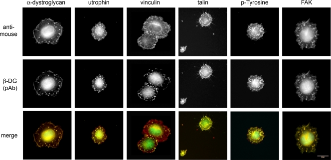
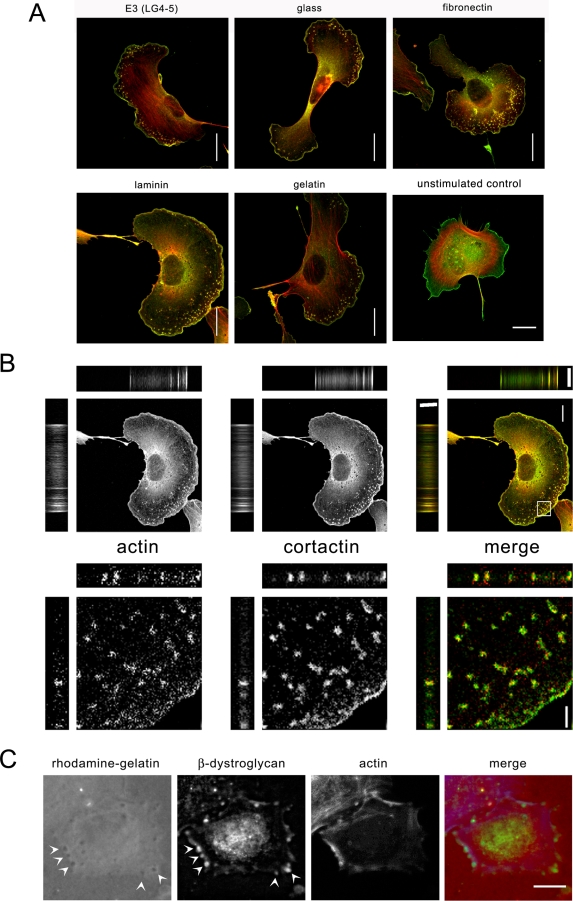
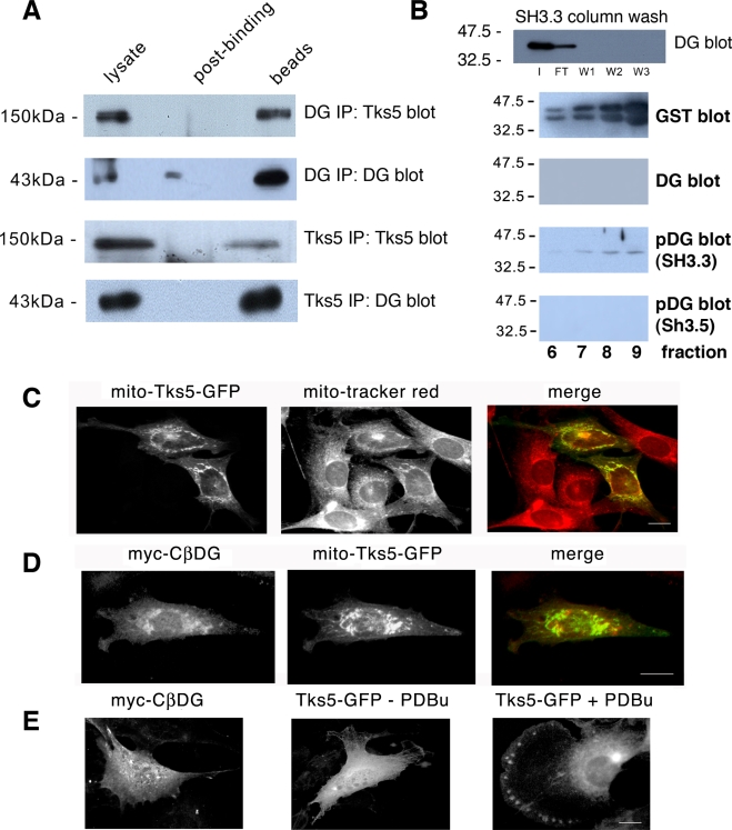
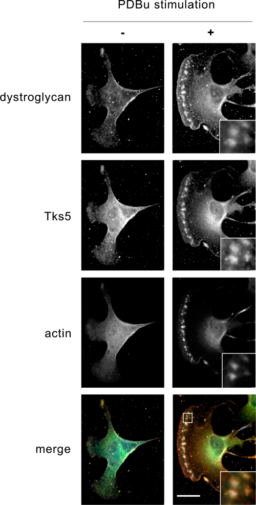
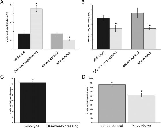
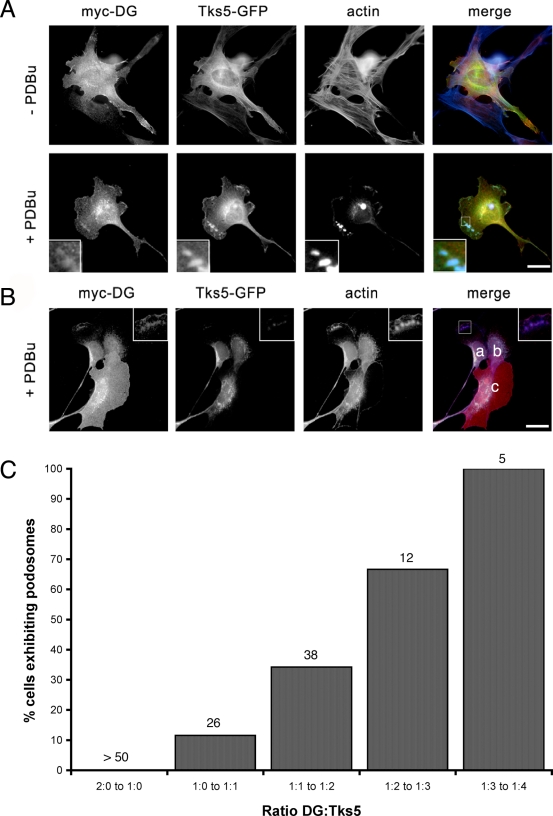
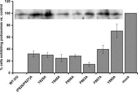
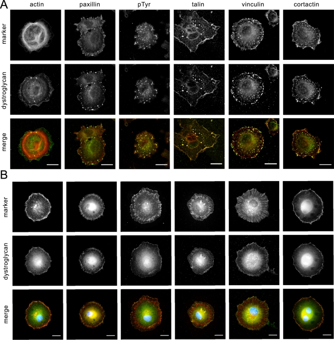
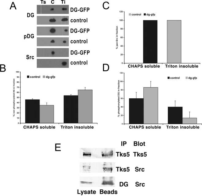
Principal findings: We have examined the role of dystroglycan in the early stages of myoblast adhesion and spreading and found that dystroglycan initially associates with other adhesion proteins in large puncta morphologically similar to podosomes. Using a human SH3 domain phage display library we identified Tks5, a key regulator of podosomes, as interacting with beta-dystroglycan. We verified the interaction by immunoprecipitation, GST-pulldown and immunfluorescence localisation. Both proteins localise to puncta during early phases of spreading, but importantly following stimulation with phorbol ester, also localise to structures indistinguishable from podosomes. Dystroglycan overexpression inhibited podosome formation by sequestering Tks5 and Src. Mutation of dystroglycan tyrosine 890, previously identified as a Src substrate, restored podosome formation.

Conclusions: We propose therefore, that Src-dependent phosphorylation of beta-dystroglycan results in the formation of a Src/dystroglycan complex that drives the SH3-mediated association between dystroglycan and Tks5 which together regulate podosome formation in myoblasts.


Conflict of interest statement

Competing Interests: The authors have declared that no competing interests exist.

Figures

Figure 1
Figure 1. Spreading myoblasts form DG rich adhesion puncta.
Myoblast cells were seeded onto tissue culture plastic and allowed to spread for one hour before fixation and staining for β-dystroglycan, and counterstained with one of the indicated cell adhesion proteins/markers. In all cases β-DG was colocalised in dense puncta either peripheral or more central to the cell, reminiscent of podosomes. Both α-dystroglycan and utrophin, components of a recognised adhesion structure, co-localised with β-dystroglycan. Other markers of classical focal adhesions such as vinculin, talin, FAK and the generic marker phospho-tyrosine also marked the β-dystroglycan containing puncta. Vinculin also clearly labelled peripheral focal adhesions, distinguishing them from the β-dystroglycan puncta. Scale bars 20 µm.
Figure 2
Figure 2. Myoblasts form podosomes in response to PDBu.
To determine if myoblast cells could form podosomes, myoblast cells were allowed to adhere and spread on various substrates, and stimulated with PDBu for 30 min and fixed and stained for the podosome markers cortactin (green) and F-actin (red). Myoblasts formed peripheral actin and cortactin containing puncta on all substrates tested. An unstimulated cell grown on a glass coverslip is also shown for comparison (A, bottom right panel). These structures are morphologically indistinguishable from podosomes (A). The structures appeared columnar within the cell and cortactin was localised around a more central actin core as seen in the Z section of the zoomed region of B. The identity of the DG and actin-rich puncta as podosomes/invadopodia was further substantiated by the degradation of rhodamine-gelatin beneath them, seen as darker areas of reduced rhodamine gelatin (red in merge, arrowed) that are coincident with DG localisation (green in merge, arrowed) and F-actin (blue in merge). Scale bars 20 µm in A and 10 µm in B, C, 2 µm in zoomed region of B.
Figure 3
Figure 3. Interaction of DG with Tks5.
A. β-DG (43 kDa) was co-immunoprecipitated from myoblast extracts with Tks5 antiserum and Tks5 (150 kDa) was co-immunoprecipitated with β-DG antibodies. Affinity purification of β-DG from myoblast extracts using the 3rd and 5th SH3 domains of Tks5 fused to GST revealed a specific interaction with only the 3rd SH3 domain (B). Western blots show peak fractions eluting from the GST-SH3 column, furthermore the β-DG recovered from the GST-SH3 column was preferentially recognised by antibodies that recognise the tyrosine phosphorylated form of β-DG (pDG) and not the non-phosphorylated form (DG). A GFP-tagged Tks5 construct (mito-Tks5-GFP; C) that was targeted to the mitochondria in myoblast cells, was also able to recruit a myc-tagged β-DG cytoplasmic domain (myc-CβDG) to the mitochondria (D) indicating an association between the two proteins in a cellular context. Both myc-CβDG is diffusely distributed when expressed alone as is Tks5-GFP in the absence of PDBu, but in the presence of PDBu clearly targets to podosome structures (E). Scale bars 10 µm.
Figure 4
Figure 4. DG and Tks5 localise to podosomes in myoblasts.
Myoblast cells, untreated (−) and following stimulation with PDBu for 30 min (+) were stained for endogenous β-DG (red), Tks5 (green) and F-actin (blue). Unstimulated cells exhibit only faint β-DG and Tks5 localisation toward the periphery in cellular protrusions, potentially in podosome cluster precursors . Following stimulation, β-DG and Tks5 were clearly localised along with F-actin to podosome structures following stimulation. The actin staining can be seen clearly to form a more central core to the podosome with Tks5 and dystroglycan around the periphery (inset). Scale bar 20 µm.
Figure 5
Figure 5. DG over-expression abolishes podosome formation.
Quantification of dystroglycan and Tks5 protein levels in control myoblast cells (wild-type), cells stably expressing GFP-tagged full-length DG (DG-GFP), shRNA against DG (knockdown) or a control shRNA (sense). Dystroglycan levels for the four cell lines are shown in A, with corresponding Tks5 levels in B. Whilst dystroglycan levels were increased by 2.3-flod and decreased by half in overexpressing and knockdown cells respectively, Tks5 levels were reduced by approximately 33% in both cases. Wild-type, sense, knockdown and myoblasts transiently expressing a myc-tagged DG construct were induced to form podosomes with PDBu and the number of cells exhibiting podosomes counted following fixation and staining. DG depletion caused a modest reduction in cells with podosomes compared to control, whereas DG over-expression completely abolished podosome formation. (C,D; mean±SEM).
Figure 6
Figure 6. The dystroglycan/Tks5 ratio is important for podosome formation.
Wild-type myoblast cells were co-transfected with myc-DG and Tks5-GFP and induced to form podosomes by stimulation with PDBu. In cells co-expressing both myc-DG and Tks5-GFP, podosomes were only formed when Tks5 expression was high, as judged by myc staining relative to GFP fluorescence; relative fluorescence ratio DG∶Tks5 1∶0.59 (A lower panels). However when myc-DG staining (red) predominated over GFP, podosomes were not formed in response to PDBU. B. cells marked in the merged image have DG∶Tks5 ratios of a. 1∶0.6; b, 1∶0.47; c, 1∶0.2. Cells are also stained for F-actin (blue). Scale bars are 10 µm. Quantitative analysis of dystroglycan-myc and Tks5-GFP ratios in transiently transfected cells reveal a ratio of Tks5∶DG that is permissive for podosome formation (C). Numbers above each column refer to numbers of cells falling within the given ratio of DG∶Tks5.
Figure 7
Figure 7. Mutation of the DG phosphorylation site restores podosome formation.
Myoblast cells were transiently transfected with dystroglycan-GFP wild-type sequence, or containing the indicated point mutation in the cytoplasmic domain. Wild-type dystroglycan fully suppressed podosome formation, whereas all mutant constructs partially suppressed the inhibition of podosomes by DG over-expression by around 20–30%, the Y890A mutation however, suppressed the inhibitory effect by over 70% compared to mock transfected controls. (mean±SEM). Inset shows typical expression levels of the dystroglycan-GFP and mutants as judged by anti-GFP western blot from a representative transient expression.
Figure 8
Figure 8. Src inhibition prevents dystroglycan localisation/podosome formation.
Myoblast cells were seeded onto tissue culture plastic and allowed to spread for one hour before fixation and staining for β-dystroglycan, F-actin and the indicated markers of cell adhesion. In A cells were untreated and formed dystroglycan-containing puncta as before. Cells in B, were treated with the Src Inhibitor PP2 (10 µM) for 1 hour before and during re-seeding. No dystroglycan-containing puncta were formed in these cells. Focal adhesions appeared to form as normal, as judged by staining for paxillin, talin, vinculin and phospho-tyrosine (pTyr). Dystroglycan staining in green in all merged images. Scale bars 20 µm.
Figure 9
Figure 9. Dystroglycan overexpression alters Src distribution.
A. A representative blot of fractionation of DG, pDG and Src in control or DG-GFP overexpressing cells; Ts, Triton soluble wash fraction; C, CHAPS soluble cytoplasmic/membrane fraction; Ti, Triton insoluble cytoskeletal fraction. Quantification from 3 independent fractionation experiments of DG (B), pDG (D) and pan Src (C) in control or DG-GFP overexpressing cells revealed an increase in pDG relative to total DG, and in the CGAPS soluble fraction relative to Triton insoluble cytoskeletal fraction, of DG overexpressing cells (B,D) Most dramatic and unexpected is a complete shift of Src from Triton insoluble cytoskeletal fraction to CHAPS soluble membrane fraction on DG overexpression (C). Immunoprecipitation from myoblast lysates with Tks5 or DG antibodies both revealed the presence of Src in the precipitated fraction of both suggesting the presence of a DG/Src/Tks5 complex in myoblast cells (E).

References

    1. Anderson C, Winder SJ, Borycki A-G. Dystroglycan protein distribution coincides with basement membranes and muscle differentiation during mouse embryogenesis. Dev Dyn. 2007;236:2627–2635. - PubMed
    1. Linder S. The matrix corroded: podosomes and invadopodia in extracellular matrix degradation. Trends in Cell Biology. 2007;17:107–117. - PubMed
    1. Tarone G, Cirillo D, Giancotti FG, Comoglio PM, Marchisio PC. Rous sarcoma virus-transformed fibroblasts adhere primarily at discrete protrusions of the ventral membrane called podosomes. Exp Cell Res. 1985;159:141–157. - PubMed
    1. Zhang D, Udagawa N, Nakamura I, Murakami H, Saito S, et al. The small GTP-binding protein, rho p21, is involved in bone resorption by regulating cytoskeletal organization in osteoclasts. J Cell Sci. 1995;108:2285–2292. - PubMed
    1. Linder S, Nelson D, Weiss M, Aepfelbacher M. Wiskott-Aldrich syndrome protein regulates podosomes in primary human macrophages. Proc Nat Acad Sci US. 1999;96:9648–9653. - PMC - PubMed

Publication types

MeSH terms