. 2008 Jul 7;2(1):64-71.
doi: 10.3389/neuro.01.001.2008. eCollection 2008 Jul.

Layer, column and cell-type specific genetic manipulation in mouse barrel cortex

Affiliations

Layer, column and cell-type specific genetic manipulation in mouse barrel cortex

Rachel Aronoff et al. Front Neurosci. .

Abstract

Sensory information is processed in distributed neuronal networks connected by intricate synaptic circuits. Studies of the rodent brain can provide insight into synaptic mechanisms of sensory perception and associative learning. In particular, the mouse whisker sensorimotor system has recently begun to be investigated through combinations of imaging and electrophysiology, providing data correlating neural activity with behaviour. In order to go beyond such correlative studies and to pinpoint the contributions of individual genes to brain function, it is critical to make highly controlled and specific manipulations. Here, we review recent progress towards genetic manipulation of targeted genes in specific neuronal cell types located in a selected cortical layer of a well-defined cortical column of mouse barrel cortex. The unprecedented precision of such genetic manipulation within highly specific neural circuits may contribute significantly to progress in understanding the molecular and synaptic determinants of simple forms of sensory perception and associative learning.

Keywords: NMDA receptors; barrel cortex; genetic manipulation; lentivirus.


Figures

Figure 1
Figure 1
The barrel cortex. (A) An impressive array of whiskers on the snout of the rodent sends sensory information to the primary somatosensory barrel cortex (S1) via the brainstem and the thalamus. The barrel cortex signals to motor cortex (M1), which in turn regulates whisker movements. Active processing of tactile whisker information therefore appears to be an important feature of this sensory pathway. (B) The layout of the whiskers (left) is precisely matched by the layout of the barrels (right) in primary somatosensory cortex. Mice and rats have the same layout of whiskers and a standard nomenclature has been developed. The C2 whisker and barrel are highlighted in yellow. (C) A barrel column is arranged in different layers. Single whisker sensory information from ventral posterior medial (VPM) thalamus arrives predominantly in a single layer 4 barrel. The supragranular layer 2/3 and the infragranular layers 5/6 are thought to perform integrative functions. Modified and reproduced with permission from Petersen (2007). Copyright from Neuron, Cell Press.
Figure 2
Figure 2
Layer and cell-type specific genetic manipulation of the barrel cortex. (A) Lentivirus can be injected directly into the mouse barrel cortex using very fine glass micropipettes. (B) Lentivector injections result in highly localized gene transduction, limited to ∼200 μm around the injection site. Injection of GFP-expressing lentivector into layer 2/3, therefore results in highly specific GFP expression in layer 2/3 and not elsewhere (left). GFP expression in pyramidal neurons is driven by the αCaMKII promoter (right). Modified and reproduced with permission from Aronoff and Petersen (2007). Frontiers in Integrative Neuroscience.
Figure 3
Figure 3
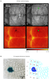
Column specific genetic manipulation of the barrel cortex. (A) Intrinsic optical imaging through the intact mouse skull can be used to non-invasively map the neocortex. Deflection of the C2 whisker evokes a localized hemodynamic signal highlighted by a green dot. The functional mapping can be related to the blood vessel layout allowing targeted craniotomy and lentivector injection. (B) Injection of lentivector was targeted to the C2 barrel column through intrinsic optical imaging. The lentivector expressed Cre recombinase and was injected into a ROSA26R Cre-reporter mouse, which expresses LacZ in the presence of Cre-activity. Modified and reproduced with permission from Aronoff and Petersen (2007). Frontiers in Integrative Neuroscience.
Figure 4
Figure 4
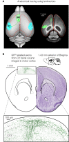
Axonal tracing using lentivectors. (A) Targeted injection of GFP expressing lentivectors into the mouse C2 barrel column, allows the analysis of the long-range axonal output. A prominent innervation of motor cortex is visualized in horizontal brain slices. (B) GFP labelled axons originating from pyramidal neurons in the C2 barrel column are shown in a coronal section of motor cortex (∼1.4 mm anterior of Bregma). Panel A modified and reproduced with permission from Ferezou et al. (2007). Copyright from Neuron, Cell Press. Panel B (right) is modified and reproduced with permission from Paxinos and Franklin (2001) The mouse brain in stereotaxic coordinates. Copyright from Academic Press.
Figure 5
Figure 5
Highly controlled knockout of NMDA receptors in the barrel cortex. (A) Imaging by two-photon microscopy of lentivector transduced pyramidal neurons expressing GFP and Cre recombinase in a floxed NR1 mouse. The Dodt contrast image (upper left), shows the whole-cell recording electrode filled with Alex 594 (upper right) targeted to a GFP expressing neuron (lower left, overlay lower right). (B) Whole-cell recording of a control layer 2/3 pyramidal neuron near the lentivector injection site. The neuron did not express GFP and had normal NMDA receptor dependent currents evoked by an extracellular stimulus in layer 4 and measured at +40 mV. (C) Whole-cell recording of a layer 2/3 pyramidal neuron expressing GFP and Cre recombinase in a floxed NR1 mouse. Synaptically evoked currents lack the long-lasting NMDA-receptor dependent component at +40 mV, indicating a functional knockout of NR1. Modified and reproduced with permission from Aronoff and Petersen (2007). Frontiers in Integrative Neuroscience.

Similar articles

Cited by

References

    1. Accolla R., Bathellier B., Petersen C. C. H., Carleton A. (2007). Differential spatial representation of taste modalities in the rat gustatory cortex. J. Neurosci. 27, 1396–140410.1523/JNEUROSCI.5188-06.2007 - DOI - PMC - PubMed
    1. Aronoff R., Petersen C. C. H. (2006). Controlled and localized genetic manipulation in the brain. J. Cell. Mol. Med. 10, 333–35210.1111/j.1582-4934.2006.tb00403.x - DOI - PMC - PubMed
    1. Aronoff R., Petersen C. C. H. (2007). Layer- and column-specific knockout of NMDA receptors in pyramidal neurons of the mouse barrel cortex. Front. Integr. Neurosci. 1, 1 10.3389/neuro.07.001.2007 - DOI - PMC - PubMed
    1. Bender V. A., Bender K. J., Brasier D. J., Feldman D. E. (2006). Two coincidence detectors for spike timing-dependent plasticity in somatosensory cortex. J. Neurosci. 26, 4166–417710.1523/JNEUROSCI.0176-06.2006 - DOI - PMC - PubMed
    1. Binzegger T., Douglas R. J., Martin K. A. (2004). A quantitative map of the circuit of cat primary visual cortex. J. Neurosci. 24, 8441–845310.1523/JNEUROSCI.1400-04.2004 - DOI - PMC - PubMed

LinkOut - more resources