Review
. 2008 Nov;118(11):3522-30.
doi: 10.1172/JCI36150.

Macrophage diversity in renal injury and repair

Affiliations
Review

Macrophage diversity in renal injury and repair

Sharon D Ricardo et al. J Clin Invest. 2008 Nov.

Abstract

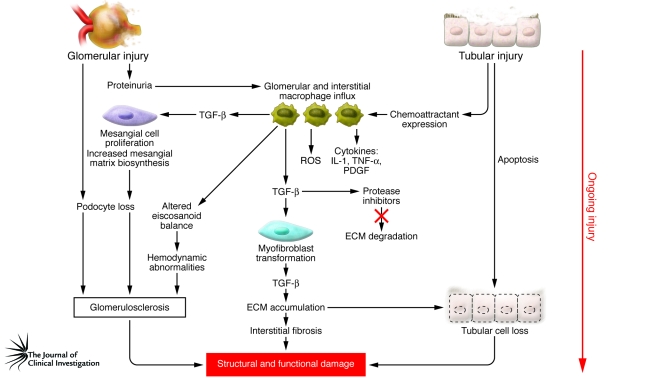
Monocyte-derived macrophages can determine the outcome of the immune response and whether this response contributes to tissue repair or mediates tissue destruction. In addition to their important role in immune-mediated renal disease and host defense, macrophages play a fundamental role in tissue remodeling during embryonic development, acquired kidney disease, and renal allograft responses. This review summarizes macrophage phenotype and function in the orchestration of kidney repair and replacement of specialized renal cells following injury. Recent advances in our understanding of macrophage heterogeneity in response to their microenvironment raise new and exciting therapeutic possibilities to attenuate or conceivably reverse progressive renal disease in the context of fibrosis. Furthermore, parallels with pathological processes in many other organs also exist.


Figures

Figure 1
Figure 1. The relationships between infiltrating macrophages and macrophage-derived products in chronic ongoing inflammation lead to structural and functional renal damage.
In response to tubular and glomerular injury/dysfunction, macrophage chemoattractants and proteinuria promote the infiltration of renal macrophages, leading to the generation of proinflammatory cytokines, vasoactive eicosanoids, and ROS. The initial injury and proinflammatory state may lead to podocyte and tubular cell apoptosis. Overproduction of TGF-β by macrophages, myofibroblasts, and mesangial cells promotes increased synthesis of glomerular and interstitial ECM proteins and decreased matrix turnover due to the synthesis of matrix-degrading protease inhibitors. The net effect of interstitial fibrosis and/or glomerulosclerosis and podocyte and tubular cell loss is the disruption to tissue architecture and loss of renal function.
Figure 2
Figure 2. Macrophage phenotype and function are critical determinants of fibrotic scarring or resolution of injury.
Monocytes from the circulation that enter the kidney in response to inflammatory cues undergo distinctive pathways of differentiation into classically activated M1 macrophages or the alternative M2 phenotype. Activation of M1 inflammatory macrophages by classical immune pathways may lead to the expression of MHC class II antigens and release of proinflammatory cytokines. In response to ongoing injury, M1 macrophages propagate inflammation and ultimately the development of fibrosis. Dependent on microenvironmental cues, M2 macrophages may be recruited from the circulation or activated in situ as a result of an M1-to-M2 phenotype switch. M2 antiinflammatory macrophages secrete regenerative trophic factors that promote cell proliferation and reduce apoptosis and stimulate angiogenesis. Macrophages derived from engrafting bone marrow myeloid progenitors may contribute to the repopulation of injured tubular epithelial and glomerular cells by a process of transdifferentiation or cell-cell fusion, leading to replacement of damaged cells. Ex vivo modulation of macrophages to form an M2 phenotype for transplantation may be used therapeutically to suppress the immune response and promote tissue remodeling, leading to structural repair and functional recovery. R, receptor.
Figure 3
Figure 3. Future therapeutic possibilities for kidney disease.
(A) Macrophage-based cellular therapies or therapeutic interventions can be envisaged that capitalize on the specialized macrophage secretome that determines differential function. Monocytes might be manipulated ex vivo to migrate to a damaged kidney, where they are preferentially M2 polarized, perhaps by inducing receptors to specific chemokines or chemoattractant molecules. Alternatively M2-type macrophages generated ex vivo from peripheral blood monocytes can be administered. Renal dendritic cells and unpolarized macrophages (M0) might also be skewed to an M2 phenotype by therapeutic manipulation of intrarenal molecular signals, such as specific cytokines, chemokines, or ECM proteins, known to direct this process in situ. (B) As more is learned about which soluble secreted macrophage products are associated with renal injury versus repair, single agents or, more likely, a cocktail of biological agents, drugs, and/or small molecules, might be administered to direct tissue recovery. This might include targeting of secondary intracellular signaling cascades that are activated by specific macrophage-derived products.

References

    1. Pistole T.G., Britko J.L. Bactericidal activity of amebocytes from the horseshoe crab, Limulus polyphemus. J. Invertebr. Pathol. 1978;31:376–382. doi: 10.1016/0022-2011(78)90233-1. - DOI - PubMed
    1. Takahashi K., Naito M. Development, differentiation, and proliferation of macrophages in the rat yolk sac. Tissue Cell. 1993;25:351–362. doi: 10.1016/0040-8166(93)90077-X. - DOI - PubMed
    1. Takahashi K., Yamamura F., Naito M. Differentiation, maturation, and proliferation of macrophages in the mouse yolk sac: a light-microscopic, enzyme-cytochemical, immunohistochemical, and ultrastructural study. J. Leukoc. Biol. 1989;45:87–96. - PubMed
    1. Morioka Y., Naito M., Sato T., Takahashi K. Immunophenotypic and ultrastructural heterogeneity of macrophage differentiation in bone marrow and fetal hematopoiesis of mouse in vitro and in vivo. J. Leukoc. Biol. 1994;55:642–651. - PubMed
    1. Hume D.A. The mononuclear phagocyte system. Curr. Opin. Immunol. 2006;18:49–53. doi: 10.1016/j.coi.2005.11.008. - DOI - PubMed

Publication types