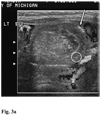
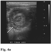
Intramural and subserosal echogenic foci on US in large-bowel intussusceptions: prognostic indicator for reducibility?
- PMID: 18982323
- PMCID: PMC2717037
- DOI: 10.1007/s00247-008-1039-y
Intramural and subserosal echogenic foci on US in large-bowel intussusceptions: prognostic indicator for reducibility?
Abstract
Background: In large-bowel intussusceptions, several US signs are known to indicate a lower likelihood of reducibility by enema. US can demonstrate echogenic dots or lines (foci) in the bowel wall, which might indicate an ischemic bowel.
Objective: To determine the presence of echogenic intramural and subserosal foci in large-bowel intussusceptions and to evaluate the degree of correlation with reducibility.
Materials and methods: Between 2001 and 2008, 74 consecutive US examinations were retrospectively evaluated by two pediatric radiologists for intramural and subserosal echogenic foci, or trapped gas, in the intussusception. The degree of correlation between the sonographic findings and reducibility was evaluated.
Results: Of 73 intussusceptions examined by US, 56 (76%) were reducible and 17 (23%) were not reducible. Out of 10 intussusceptions with intramural gas, 11 with subserosal gas, and 14 with intramural and subserosal gas, 8 (80%), 6 (56%), 9 (64%), respectively, were not reducible. The presence of intramural gas or subserosal gas or both predicted a lower chance of reduction, but with regard to the effect of these findings together, intramural gas was the only significant predictor.
Conclusion: Having intramural gas in large-bowel intussusception significantly decreases the chance of reduction.
Figures


References
-
- Bowerman RA, Silver TM, Jaffe MH. Real-time ultrasound diagnosis of intussusception in children. Radiology. 1982;143:527–529. - PubMed
-
- Swischuk LE, Hayden CK, Boulden T. Intussusception: indications for ultrasonography and an explanation of the doughnut and pseudokidney signs. Pediatr Radiol. 1985;15:388–391. - PubMed
-
- del-Pozo G, Gonzalez-Spinola J, Gomez-Anson B, et al. Intussusception: trapped peritoneal fluid detected with US--relationship to reducibility and ischemia. Radiology. 1996;201:379–383. - PubMed
-
- Hanquinet S, Anooshiravani M, Vunda A, et al. Reliability of color Doppler and power Doppler sonography in the evaluation of intussuscepted bowel viability. Pediatric surgery international. 1998;13:360–362. - PubMed
-
- Koumanidou C, Vakaki M, Pitsoulakis G, et al. Sonographic detection of lymph nodes in the intussusception of infants and young children: clinical evaluation and hydrostatic reduction. AJR Am J Roentgenol. 2002;178:445–450. - PubMed

