. 2009 Jan 9;385(1):212-25.
doi: 10.1016/j.jmb.2008.10.045. Epub 2008 Nov 1.

Differential activities of cellular and viral macro domain proteins in binding of ADP-ribose metabolites

Affiliations

Differential activities of cellular and viral macro domain proteins in binding of ADP-ribose metabolites

Maarit Neuvonen et al. J Mol Biol. .

Abstract

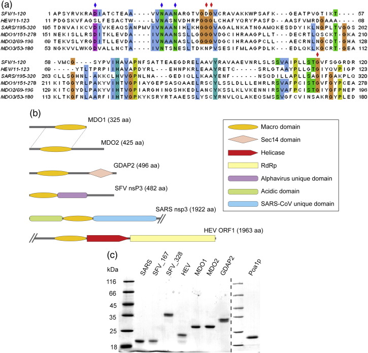
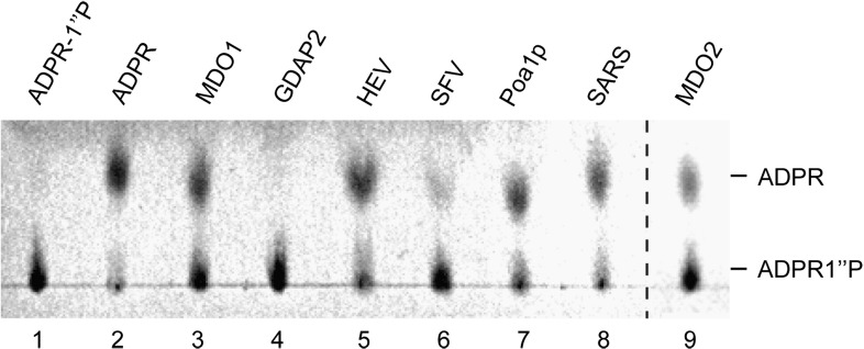
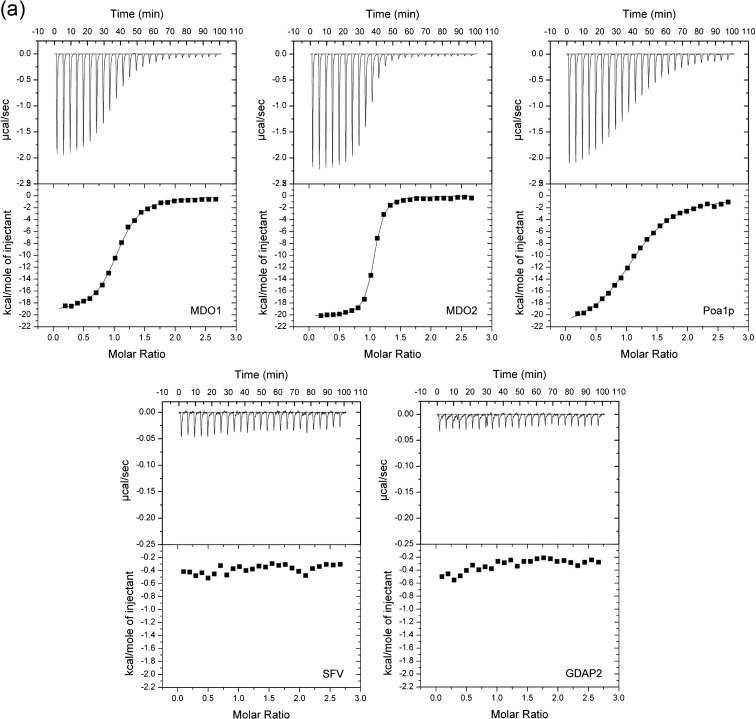
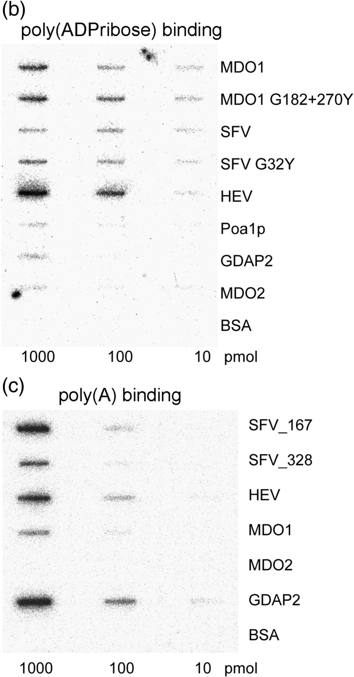
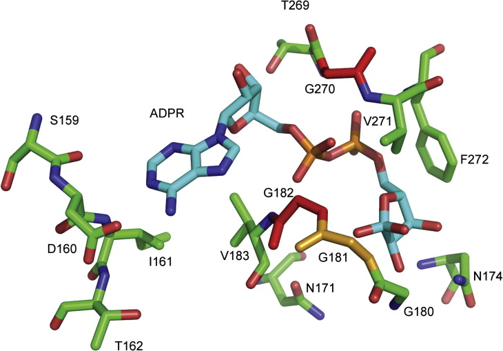
Macro domain is a highly conserved protein domain found in both eukaryotes and prokaryotes. Macro domains are also encoded by a set of positive-strand RNA viruses that replicate in the cytoplasm of animal cells, including coronaviruses and alphaviruses. The functions of the macro domain are poorly understood, but it has been suggested to be an ADP-ribose-binding module. We have here characterized three novel human macro domain proteins that were found to reside either in the cytoplasm and nucleus [macro domain protein 2 (MDO2) and ganglioside-induced differentiation-associated protein 2] or in mitochondria [macro domain protein 1 (MDO1)], and compared them with viral macro domains from Semliki Forest virus, hepatitis E virus, and severe acute respiratory syndrome coronavirus, and with a yeast macro protein, Poa1p. MDO2 specifically bound monomeric ADP-ribose with a high affinity (K(d)=0.15 microM), but did not bind poly(ADP-ribose) efficiently. MDO2 also hydrolyzed ADP-ribose-1'' phosphate, resembling Poa1p in all these properties. Ganglioside-induced differentiation-associated protein 2 did not show affinity for ADP-ribose or its derivatives, but instead bound poly(A). MDO1 was generally active in these reactions, including poly(A) binding. Individual point mutations in MDO1 abolished monomeric ADP-ribose binding, but not poly(ADP-ribose) binding; in poly(ADP-ribose) binding assays, the monomer did not compete against polymer binding. The viral macro proteins bound poly(ADP-ribose) and poly(A), but had a low affinity for monomeric ADP-ribose. Thus, the viral proteins do not closely resemble any of the human proteins in their biochemical functions. The differential activity profiles of the human proteins implicate them in different cellular pathways, some of which may involve RNA rather than ADP-ribose derivatives.


Figures

Fig. 1
Fig. 1
Macro domain proteins of human and viral origin. (a) Multiple amino acid sequence alignment of macro domain proteins derived from SFV, HEV, and SARS-CoV with human MDO1, MDO2, and GDAP2 (MDO3). Colored bars highlight conserved amino acids with over 51% identity between the aligned proteins. Blue diamonds on top of the sequence indicate the amino acids that were mutated in SFV, and red diamonds indicate the amino acids that were mutated both in SFV and in MDO1. (b) Domain structures of human MDO1, MDO2, and GDAP2, and viral macro proteins from SFV, HEV, and SARS-CoV. Macro-domain-containing proteins from SARS-CoV and HEV are shown only partially, as indicated by slashes, but their full lengths are given by the numbers in parentheses. The region with a high level of homology between MDO1 and MDO2 is indicated by dotted lines. Predicted domains are indicated by colored symbols. RdRp, RNA-dependent RNA polymerase. (c) Purified macro domain proteins were analyzed by SDS-PAGE on 12% gels followed by staining with Coomassie blue. Lanes from left to right: Molecular mass marker, SARS-CoV nsp3 (174 aa), SFV nsP3 (167 aa), SFV nsP3 (328 aa), HEV macro (185 aa), MDO1 (243 aa), MDO2 (243 aa), GDAP2 (231 aa), molecular mass marker, and Poa1p (177 aa). The lengths of the proteins given above exclude the 13-aa to 17-aa N-terminal vector-derived sequence.
Fig. 2
Fig. 2
ADPR-1″P phosphatase activity of macro domains. The indicated macro domain proteins were incubated with ADPR-1″P for 1 h at 28 °C, and reaction products were analyzed by TLC and visualized under UV illumination. Lanes 1 and 2 contain the controls as indicated: ADPR-1″P incubated without the addition of protein, and pure ADP-ribose.
Fig. 3
Fig. 3
Macro domain proteins of different origins have different affinities for ADP-ribose monomer, poly(ADP-ribose), and poly(A). (a) Isothermal titration calorimetry of ADP-ribose binding. Titration curves show the stepwise addition of ADP-ribose into a solution containing the purified MDO1, MDO2, Poa1p, SFV, and GDAP2 macro domains in the presence of 100 mM NaCl (500 mM NaCl for SFV macro) at 30 °C. The lower parts of the figures show the fit of the measured data (first data point omitted) to an equilibrium binding isotherm. The protein assayed is indicated on the bottom-right corner of each panel. (b) Binding of 32P-labeled poly(ADP-ribose) by macro domain proteins blotted onto a nitrocellulose membrane. About 10 pmol, 100 pmol, or 1000 pmol of each protein was immobilized onto the membrane, followed by blocking with milk proteins. After 1 h of incubation with [32P]poly(ADP-ribose), the membrane was washed carefully, and bound radioactivity was detected with PhosphorImager. Bovine serum albumin was used as negative control. In addition to the wild-type macro proteins, a double mutant (G182Y + G270Y) of MDO1 and a single mutant (G32Y) of SFV nsP3 are illustrated. (c) Binding of 32P-labeled poly(A) by macro proteins was tested similarly to the poly(ADP-ribose) binding experiment.
Fig. 3
Fig. 3
Macro domain proteins of different origins have different affinities for ADP-ribose monomer, poly(ADP-ribose), and poly(A). (a) Isothermal titration calorimetry of ADP-ribose binding. Titration curves show the stepwise addition of ADP-ribose into a solution containing the purified MDO1, MDO2, Poa1p, SFV, and GDAP2 macro domains in the presence of 100 mM NaCl (500 mM NaCl for SFV macro) at 30 °C. The lower parts of the figures show the fit of the measured data (first data point omitted) to an equilibrium binding isotherm. The protein assayed is indicated on the bottom-right corner of each panel. (b) Binding of 32P-labeled poly(ADP-ribose) by macro domain proteins blotted onto a nitrocellulose membrane. About 10 pmol, 100 pmol, or 1000 pmol of each protein was immobilized onto the membrane, followed by blocking with milk proteins. After 1 h of incubation with [32P]poly(ADP-ribose), the membrane was washed carefully, and bound radioactivity was detected with PhosphorImager. Bovine serum albumin was used as negative control. In addition to the wild-type macro proteins, a double mutant (G182Y + G270Y) of MDO1 and a single mutant (G32Y) of SFV nsP3 are illustrated. (c) Binding of 32P-labeled poly(A) by macro proteins was tested similarly to the poly(ADP-ribose) binding experiment.
Fig. 4
Fig. 4
Structure prediction of the MDO1 ligand binding site. The ADP-ribose bound to the MDO1 ligand-binding pocket is shown as predicted, using SARS-CoV macro domain as model. Sites of mutations inactivating ADP-ribose binding (residues G182 and G270) are highlighted in red, and the site of mutation reducing binding (G181) is shown in orange. In the SFV macro sequence, positions 31 and 32 correspond to MDO1 residues 181–182, and position 112 corresponds to MDO1 residue 270. The residues corresponding to the additionally mutated sites in SFV are also seen in this rendering: residues D10, N21, and N24 in the SFV sequence correspond to MDO1 D160, N171, and N174, respectively.
Fig. 5
Fig. 5
Expression and localization of the human macro proteins MDO1, MDO2, and GDAP2 in HeLa cells. (a) Western blot analysis of EGFP-tagged proteins, as detected by antibodies against EGFP. An EGFP coding vector without fusion partner was used as positive control (lane 1), and mock-transfected cells were used as negative controls (lane 8). Either the full-length protein (lanes 3, 5, and 7) or the macro domain with flanking sequences (length indicated by the numbers following the name of the protein; lanes 2, 4, and 6) was expressed, as indicated on top. Molecular mass marker is shown on the left. (b) MDO1 fused to a myc epitope was cotransfected with YFP linked to a mitochondrial localization signal (mt-YFP) and detected by indirect immunofluorescence using anti-MDO1 antibodies. Localization of mt-YFP was detected in the green channel, and anti-MDO1 staining was detected in the red channel. Colocalization of the signals is illustrated in yellow. (c) The 243-aa C-terminal of MDO1 detected by anti-MDO1 did not show mitochondrial localization. Full-length MDO2 (d) and GDAP2 (e) were expressed as C-terminally EGFP-tagged recombinant proteins in HeLa cells and were detected by EGFP fluorescence both in the cytosol and in the nucleus.

References

    1. Pehrson J.R., Fuji R.N. Evolutionary conservation of histone macroH2A subtypes and domains. Nucleic Acids Res. 1998;26:2837–2842. - PMC - PubMed
    1. Anantharaman V., Koonin E.V., Aravind L. Comparative genomics and evolution of proteins involved in RNA metabolism. Nucleic Acids Res. 2002;30:1427–1464. - PMC - PubMed
    1. Allen M.D., Buckle A.M., Cordell S.C., Löwe J., Bycroft M. The crystal structure of AF1521 a protein from Archaeoglobus fulgidus with homology to the non-histone domain of macroH2A. J. Mol. Biol. 2003;330:503–511. - PubMed
    1. Martzen M.R., McCraith S.M., Spinelli S.L., Torres F.M., Fields S., Grayhack E.J., Phizicky E.M. A biochemical genomics approach for identifying genes by the activity of their products. Science. 1999;286:1153–1155. - PubMed
    1. Karras G.I., Kustatscher G., Buhecha H.R., Allen M.D., Pugieux C., Sait F., et al. The macro domain is an ADP-ribose binding module. EMBO J. 2005;24:1911–1920. - PMC - PubMed

Publication types

MeSH terms