

Review
doi: 10.1016/j.cell.2008.10.008.
The self-tuning neuron: synaptic scaling of excitatory synapses
Affiliations
- PMID: 18984155
- PMCID: PMC2834419
- DOI: 10.1016/j.cell.2008.10.008
Item in Clipboard
Review
The self-tuning neuron: synaptic scaling of excitatory synapses
Cell.
.
Abstract
Homeostatic synaptic scaling is a form of synaptic plasticity that adjusts the strength of all of a neuron's excitatory synapses up or down to stabilize firing. Current evidence suggests that neurons detect changes in their own firing rates through a set of calcium-dependent sensors that then regulate receptor trafficking to increase or decrease the accumulation of glutamate receptors at synaptic sites. Additional mechanisms may allow local or network-wide changes in activity to be sensed through parallel pathways, generating a nested set of homeostatic mechanisms that operate over different temporal and spatial scales.
Figures

A) Correlated presynaptic and postsynaptic firing induces long-term potentiation (LTP), which then allows the presynaptic neuron to drive the postsynaptic neuron more strongly. This increases the correlation between presynaptic and postsynaptic activation, which drives more LTP, and so on in an unconstrained positive feedback cycle. B) Unconstrained LTP will lose synapse-specificity, because when one input undergoes LTP and drives the postsynaptic neuron more strongly, it makes it easier for other inputs to make the postsynaptic neuron fire, and they begin to undergo LTP as well. C) Homeostatic synaptic scaling prevents this runaway potentiation. When LTP of one input increases postsynaptic firing, synaptic scaling will reduce the strength of all synaptic inputs until the firing rate returns to control levels. Note that synaptic strengths are reduced proportionally, so that the relative strength of the potentiated synapse remains the same.
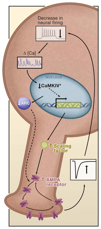
A drop in neuronal firing leads to a drop in somatic calcium. This decreases the amount of activated CaMKIV (CaMKIV*) in the nucleus, and increases transcription of a “Scaling Factor” that enhances AMPA receptor accumulation at synapses through an unknown mechanism. This increases excitatory synaptic strength and raises firing rates back to target levels. There may be several signaling pathways that can act in parallel to generate synaptic scaling (dashed arrows); for example, decreased neuronal excitation will decrease the immediate early gene product Arc; reduced Arc levels increase AMPA receptor accumulation by reducing endocytosis.
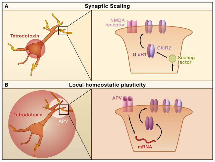
A. Blocking postsynaptic firing while leaving presynaptic and network activity intact scales up synaptic strengths in the dendrites, via a mechanism that results in increased accumulation of both GluR1 and GluR2 subunits of AMPA receptors. B. When action potential firing is blocked and NMDA receptor activation is locally blocked with the antagonist APV, there is a local increase in synaptic GluR1 accumulation that requires local dendritic protein synthesis.
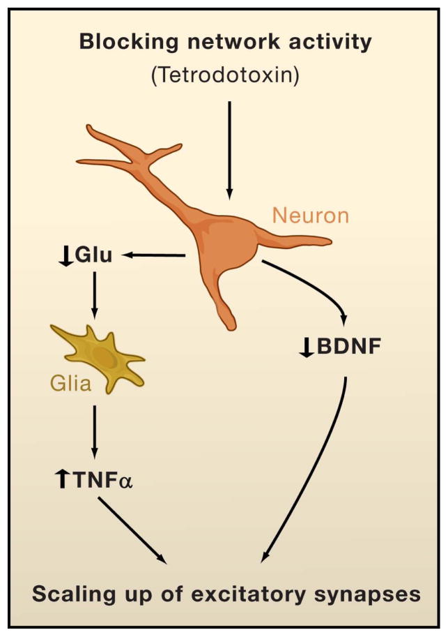
Blocking network activity with tetrodotoxin reduces release of the brain-derived neurotrophic factor (BDNF) from neurons, and the cytokine tumor necrosis factor α (TNFα) from glial cells. A chronic (several days) reduction in BDNF and increase in TNFα both scale up excitatory synapses onto pyramidal neurons.

A firing rate set-point can be constructed out of two opposing synaptic scaling factors. Factor+ (solid red line) increases in activation as activity rises, and scales down the strengths of excitatory synapses strengths. Factor- (solid blue line) increases in activation as activity falls, and scales up synaptic strengths. The firing rate at which these two opposing forces equalize is the activity set-point (solid black circle). If one of these factors is reduced or increased in magnitude, the activity set-point will shift: for example, if Factor+ is reduced in magnitude (dashed blue line), the activity set-point will shift to a more negative value (solid gray circle).
References
-
- Abbott LF, Nelson SB. Synaptic plasticity: taming the beast. Nat Neurosci. 2000;3:1178–1183. - PubMed
-
- Abraham WC, Bear MF. Metaplasticity: the plasticity of synaptic plasticity. Trends In Neurosciences. 1996;19:126–130. - PubMed
-
- Beattie EC, Stellwagen D, Morishita W, Bresnahan JC, Ha BK, Von Zastrow M, Beattie MS, Malenka RC. Control of synaptic strength by glial TNFalpha. Science. 2002;295:2282–2285. - PubMed
-
- Bessis A, Bechade C, Bernard D, Roumier A. Microglial control of neuronal death and synaptic properties. Glia. 2007;55:233–238. - PubMed
Publication types
MeSH terms
Grants and funding
LinkOut - more resources
Full Text Sources
Other Literature Sources
