. 2009 Jan 2;284(1):414-425.
doi: 10.1074/jbc.M808390200. Epub 2008 Nov 4.

Purification and characterization of the lipid A 1-phosphatase LpxE of Rhizobium leguminosarum

Affiliations

Purification and characterization of the lipid A 1-phosphatase LpxE of Rhizobium leguminosarum

Mark J Karbarz et al. J Biol Chem. .

Abstract

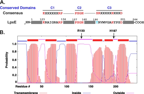
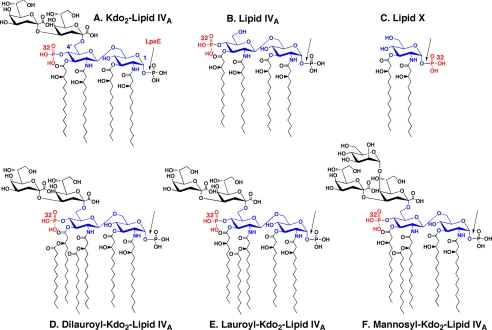
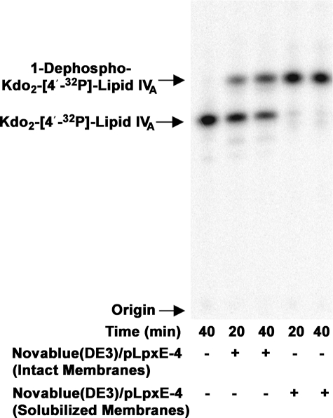
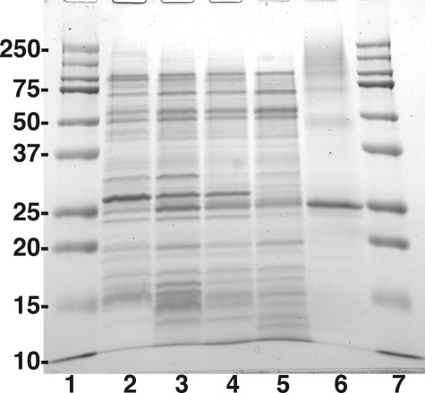
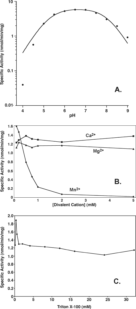
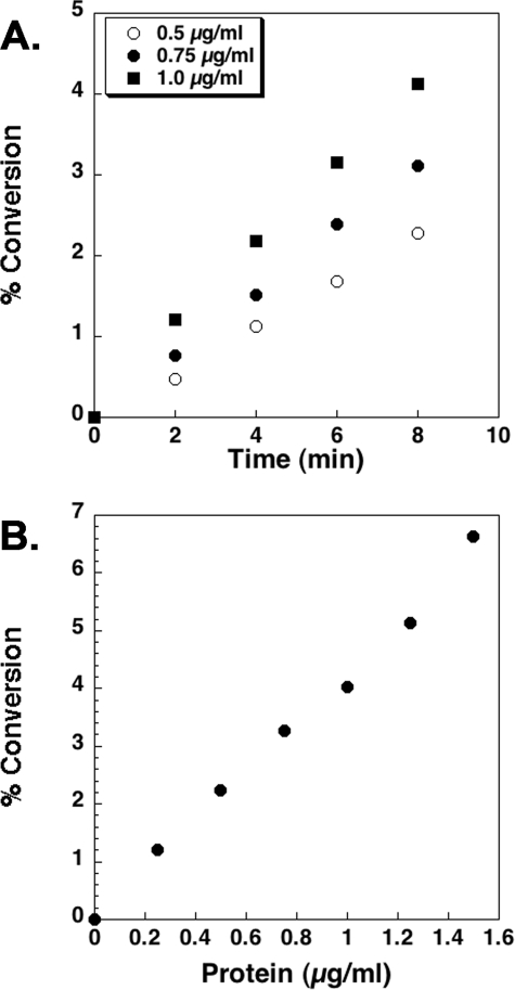
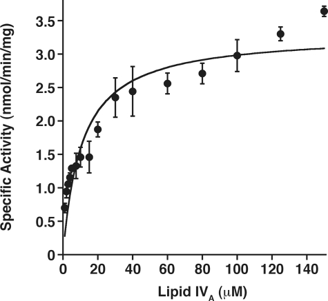
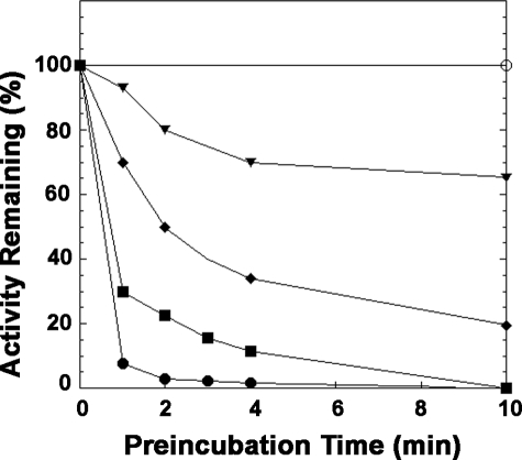
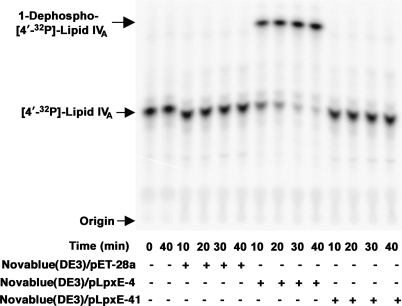
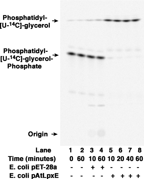
LpxE, a membrane-bound phosphatase found in Rhizobium leguminosarum and some other Gram-negative bacteria, selectively dephosphorylates the 1-position of lipid A on the outer surface of the inner membrane. LpxE belongs to the family of lipid phosphate phosphatases that contain a tripartite active site motif and six predicted transmembrane helices. Here we report the purification and characterization of R. leguminosarum LpxE. A modified lpxE gene, encoding a protein with an N-terminal His6 tag, was expressed in Escherichia coli. The protein was solubilized with Triton X-100 and purified to near-homogeneity. Gel electrophoresis reveals a molecular weight consistent with the predicted 31 kDa. LpxE activity is dependent upon Triton X-100, optimal near pH 6.5, and Mg2+-independent. The H197A and R133A substitutions inactivate LpxE, as does treatment with diethyl pyrocarbonate. In a mixed micelle assay system, the apparent Km for the precursor lipid IV(A) is 11 microm. Substrates containing the 3-deoxy-d-manno-oct-2-ulosonic acid disaccharide are dephosphorylated at similar rates to lipid IV(A), whereas glycerophospholipids like phosphatidic acid or phosphatidylglycerol phosphate are very poor substrates. However, an LpxE homologue present in Agrobacterium tumefaciens is selective for phosphatidylglycerol phosphate, demonstrating the importance of determining substrate specificity before assigning the functions of LpxE-related proteins. The availability of purified LpxE will facilitate the preparation of novel 1-dephosphorylated lipid A molecules that are not readily accessible by chemical methods.


Figures

FIGURE 1.
FIGURE 1.
LpxE active site motifs and predicted transmembrane topology. The primary sequence of native R. leguminosarum LpxE shows homology to the conserved tripartite sequence motifs of the type 2 lipid phosphate phosphatase family (panel A). For panel B, the LpxE topology was predicted using the transmembrane hidden Markov model (TMHMM) (74). All three active site domains are predicted to face the periplasm in the inner membrane, based on this model. For LpxE, domain 1 begins near the extracellular end of helix 3, extending from residues 123–134; domain 2 is located within helix 4, residues 157–160; and domain 3, residues 190–201, encompasses part of helix 5 and the small downstream extracellular loop. The arrows denote the locations of key conserved residues (Arg-133 and His-197).
FIGURE 2.
FIGURE 2.
Structures of Kdo2-lipid IVA and related substrates used to assay LpxE. The seven enzymes that make Kdo2-lipid IVA in E. coli are also found in R. leguminosarum and R. etli (28), and with the possible exception of lpxH (50, 73), the genes encoding them are present in single copy in the R. leguminosarum genome. The glucosamine residues and their numbering are shown in blue, and the bond cleaved by LpxE is indicated with the arrow. The location of the radiolabeled phosphate moiety in each substrate is indicated in red. Although most active with substrates containing a Kdo disaccharide and one secondary acyl chain, the LpxE phosphatase does not require the Kdo moiety, allowing the use of the precursor lipid IVA as a model substrate. The monosaccharide lipid A precursor lipid X is a poor substrate. Because the active site of LpxE is oriented toward the periplasmic surface of the inner membrane, however, it does not normally have access to LPS precursors lacking the Kdo moiety or the 2′ secondary acyl chain.
FIGURE 3.
FIGURE 3.
Activation of LpxE by solubilization of Novablue(DE3)/pLpxE-4 membranes with 4% Triton X-100. Membranes of Novablue(DE3)/pLpxE-4 were solubilized with 4% Triton X-100, ultracentrifuged to remove insoluble material, and assayed for 1-phosphatase activity with Kdo2-[4′-32P]lipid IVA as substrate under otherwise standard conditions at 30 °C. The no enzyme control is shown in the 1st lane. Membranes from Novablue (DE3)/pLpxE-4 that had not been solubilized with Triton X-100 were used at 0.5 mg/ml in the 2nd and 3rd lanes. Triton X-100-solubilized supernatant of Novablue(DE3)/pLpxE-4 membranes was used at 0.5 mg/ml in the 4th and 5th lanes. At each time point, a 4-μl portion was quenched, and the extent of dephosphorylation was assessed by TLC and PhosphorImager analysis.
FIGURE 4.
FIGURE 4.
Gel electrophoresis of LpxE at various stages of the purification. The molecular weight standards, shown in lanes 1 and 7, are 10, 15, 20, 25, 37, 50, 75, 100, 150, and 250 kDa; lane 2, 10 μg of membrane protein from Novablue(DE3)/pET-28a induced with IPTG; lane 3, 10 μg of membrane protein from Novablue(DE3)/pLpxE-4 induced with IPTG; lane 4, 10 μg of 4% Triton X-100-treated LpxE membranes from Novablue(DE3)/pLpxE-4 induced with IPTG (pre-centrifugation); lane 5, 10 μg of 4% Triton X-100-solubilized LpxE membranes (post-centrifugation); lane 6, 10 μg of purified LpxE protein (after the Ni2+-NTA column).
FIGURE 5.
FIGURE 5.
Effect of pH, divalent cations, and Triton X-100 on LpxE activity. The 1-phosphatase activity was measured at the indicated pH values in a uniform triple buffer system (panel A). The curve represents a fit of the pH-rate equation as described under “Experimental Procedures.” The pKa and pKb values derived from the fit are 5.25 ± 0.07 and 8.04 ± 0.07. The 1-phosphatase activity was measured in the presence of MgCl2, CaCl2, or MnCl2 (panel B) or at various concentrations of Triton X-100 (panel C) under otherwise standard conditions. The points are connected for ease of visualization. The data shown in each panel are from a single representative experiment.
FIGURE 6.
FIGURE 6.
Time and protein concentration dependence of R. leguminosarum LpxE. Dephosphorylation of [4′-32P]lipid IVA is linear with time (panel A) and protein concentration when assayed for 10 min under standard conditions at pH 6.5 (panel B). After prolonged incubation, or in the presence of high enzyme concentrations, the reaction goes to completion (data not shown). The data shown in each panel are from a single representative experiment.
FIGURE 7.
FIGURE 7.
Steady state kinetics of purified LpxE in a mixed micelle system. Standard assay conditions were used, but the lipid IVA substrate concentrations were varied. A fit of the Michaelis-Menten equation to the data gives an apparent Km of 11 ± 2 μm for lipid IVA and an apparent Vmax of 3.3 ± 0.2 nmol/min/mg. The data shown are the average of several experiments with the standard deviation shown.
FIGURE 8.
FIGURE 8.
Time course of inactivation of R. leguminosarum LpxE by DEPC. Purified LpxE (20 μm) was preincubated at room temperature with 0 (○), 10 μm (▾), 20 μm (♦), 40 μm (▪), or 80 μm (•) DEPC. At the indicated times, a portion of the preincubation mixture was quenched with imidazole, diluted, and assayed for remaining LpxE activity. The points are connected for ease of visualization. The data shown are from a single representative experiment.
FIGURE 9.
FIGURE 9.
Absence of LpxE activity of the H197A LpxE mutant. Membranes isolated from Novablue(DE3)/pET-28a (empty vector), Novablue(DE3)/pLpxE-4 (wild-type LpxE), and Novablue(DE3)/pLpxE-41 (H197A) were solubilized with 4% Triton X-100 and assayed in the presence of 0.1 mg/ml protein for 1-phosphatase activity at 30 °C.
FIGURE 10.
FIGURE 10.
The LpxE homologue of A. tumefaciens is a PGP phosphatase. The assay was carried out under standard conditions at the indicated times with no protein added (lanes 1 and 2), 0.5 mg/ml membrane protein from cells harboring either the vector control (lanes 3 and 4), or pAtLpxE (lanes 5-8).
FIGURE 11.
FIGURE 11.
Topography of the active sites of LpxE and other lipid A modification enzymes in R. etli and R. leguminosarum. The structure of the conserved intermediate Kdo2-lipid IVA is shown in Fig. 2. The evidence for the existence and orientation of these enzymes and transporters is reviewed elsewhere (2). The color scheme is as follows: glucosamine, blue; Kdo, white; galacturonic acid, cyan; aminogluconate, magenta; phosphate groups, red; fatty acyl chains, green; enzymes, red letters; proposed transport proteins, black letters. The active sites of both LpxE (the 1-phosphatase) and LpxF (the 4′-phosphatase) face the periplasm, preventing premature dephosphorylation of key precursors such as lipid IVA.

Similar articles

Cited by

References

    1. Raetz, C. R. H., and Whitfield, C. (2002) Annu. Rev. Biochem. 71 635-700 - PMC - PubMed
    1. Raetz, C. R. H., Reynolds, C. M., Trent, M. S., and Bishop, R. E. (2007) Annu. Rev. Biochem. 76 295-329 - PMC - PubMed
    1. Fahy, E., Subramaniam, S., Brown, H. A., Glass, C. K., Merrill, A. H., Jr., Murphy, R. C., Raetz, C. R. H., Russell, D. W., Seyama, Y., Shaw, W., Shimizu, T., Spener, F., van Meer, G., VanNieuwenhze, M. S., White, S. H., Witztum, J. L., and Dennis, E. A. (2005) J. Lipid Res. 46 839-862 - PubMed
    1. Nikaido, H. (2003) Microbiol. Mol. Biol. Rev. 67 593-656 - PMC - PubMed
    1. Galloway, S. M., and Raetz, C. R. H. (1990) J. Biol. Chem. 265 6394-6402 - PubMed

Publication types

MeSH terms

Substances

LinkOut - more resources