. 2008 Dec;36(22):e150.
doi: 10.1093/nar/gkn691. Epub 2008 Nov 4.

Errors in the bisulfite conversion of DNA: modulating inappropriate- and failed-conversion frequencies

Affiliations

Errors in the bisulfite conversion of DNA: modulating inappropriate- and failed-conversion frequencies

Diane P Genereux et al. Nucleic Acids Res. 2008 Dec.

Erratum in

  • Nucleic Acids Res. 2009 Aug;37(15):5235

Abstract

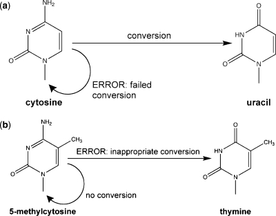
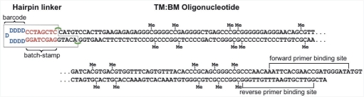
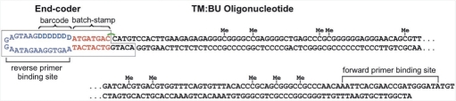
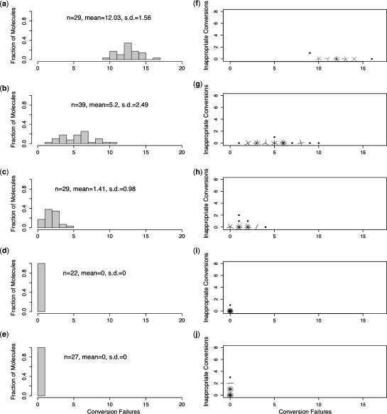
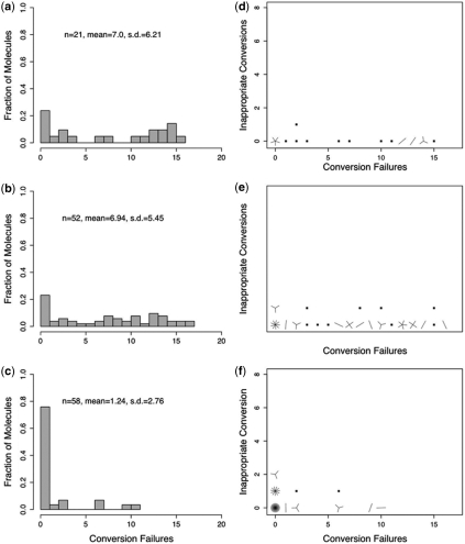
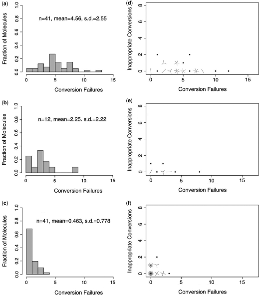
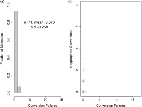
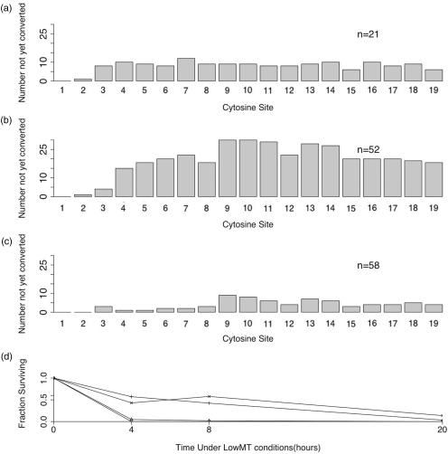
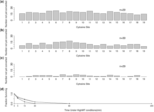
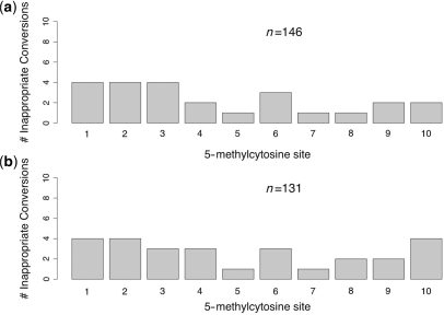
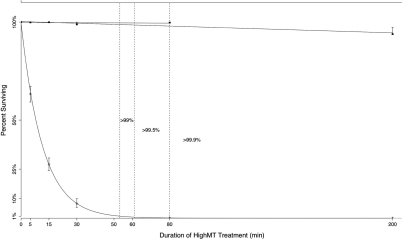
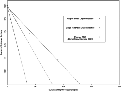
Bisulfite treatment can be used to ascertain the methylation states of individual cytosines in DNA. Ideally, bisulfite treatment deaminates unmethylated cytosines to uracils, and leaves 5-methylcytosines unchanged. Two types of bisulfite-conversion error occur: inappropriate conversion of 5-methylcytosine to thymine, and failure to convert unmethylated cytosine to uracil. Conventional bisulfite treatment requires hours of exposure to low-molarity, low-temperature bisulfite ('LowMT') and, sometimes, thermal denaturation. An alternate, high-molarity, high-temperature ('HighMT') protocol has been reported to accelerate conversion and to reduce inappropriate conversion. We used molecular encoding to obtain validated, individual-molecule data on failed- and inappropriate-conversion frequencies for LowMT and HighMT treatments of both single-stranded and hairpin-linked oligonucleotides. After accounting for bisulfite-independent error, we found that: (i) inappropriate-conversion events accrue predominantly on molecules exposed to bisulfite after they have attained complete or near-complete conversion; (ii) the HighMT treatment is preferable because it yields greater homogeneity among sites and among molecules in conversion rates, and thus yields more reliable data; (iii) different durations of bisulfite treatment will yield data appropriate to address different experimental questions; and (iv) conversion errors can be used to assess the validity of methylation data collected without the benefit of molecular encoding.


Figures

Figure 1.
Figure 1.
Responses of methylated and unmethylated cytosines to bisulfite treatment. (a) Unmethylated cytosines treated with bisulfite are either converted to uracil, or fail to be converted and remain as cytosine. (b) the 5-methylcytosines treated with bisulfite either do not undergo conversion, or are inappropriately converted to thymine.
Figure 2.
Figure 2.
A hairpin-linked TM:BM oligonucleotide. The 5-methylcytosines are present at each of 10 CpG sites on both top, TM, and bottom, BM, strands of TM:BM, and are indicated by ‘Me’. Each hairpin linker contains a batchstamp common to all molecules processed in a given experiment, and a randomly generated barcode (19). Hairpin-linked molecules have primer binding sites on the top and bottom strands of the oligonucleotide, permitting subsequent PCR amplification.
Figure 3.
Figure 3.
An end-coded TM oligonucleotide ligated while base-paired with BU. The 5-methylcytosines are present at 10 CpG sites on the top strand, TM, and are indicated with ‘Me’. The end-coder (Burden et al., manuscript in preparation) contains a batchstamp common to all molecules processed in a given experiment, and a randomly generated barcode. The end-coder is attached to the top, TM, but not the bottom, BU, strand of TM:BU. Thus, the bottom strand will separate from the top strand under denaturating conditions. Oligonucleotides labeled with end-coder have a forward primer binding site on TM, and a reverse-primer binding site of 21 nt, indicated here in purple, on the end-coder itself (after Figure 1 of Burden et al., manuscript in preparation).
Figure 4.
Figure 4.
Single-stranded oligonucleotides exposed to HighMT bisulfite. Distributions of failed-conversion counts for molecules treated in HighMT bisulfite for 5 min (a), 15 min (b), 30 min (c), 80 min (d) and 200 min (e). We use n to indicate the number of molecules examined; conversion failures are summarized by the mean number of failed-conversion events per molecule, and the SD on this mean. Sunflower plots of individual-molecule failed- and inappropriate-conversion counts for molecules treated with HighMT bisulfite for 5 min (f), 15 min (g), 30 min (h), 80 min (i) and 200 min (j). A single point indicates one molecule, a line indicates two and a z-petaled ‘flower’ indicates z-molecules.
Figure 5.
Figure 5.
Single-stranded oligonucleotides exposed to LowMT bisulfite. Distributions of failed-conversion counts for bisulfite treatments of 4 h (a), 8 h (b) and 20 h (c). Sunflower plots of individual molecule failed- and inappropriate-conversion counts for treatments of 4 h (d), 8 h (e) and 20 h (f). We use n to indicate the number of molecules examined; conversion failures are summarized by the mean number of failed-conversion events per molecule and the SD on this mean. A single point indicates one molecule, a line indicates two and a z-petaled ‘flower’ indicates z-molecules.
Figure 6.
Figure 6.
Hairpin-linked oligonucleotides exposed to HighMT bisulfite. Distributions of failed-conversion counts for bisulfite treatments of 40 min (a), 60 min (b) and 90 min (c). We use n to indicate the number of molecules examined; conversion failures are summarized by the mean number of failed-conversion events per molecule and the SD on this mean. Sunflower plots of individual-molecule failed- and inappropriate-conversion counts for bisulfite treatments of 40 min (d), 60 min (e) and 90 min (f). A single point indicates one molecule, a line indicates two and a z-petaled ‘flower’ indicates z-molecules.
Figure 7.
Figure 7.
Hairpin-linked DNA treated under the published hairpin-bisulfite PCR protocol. (a) Distribution of failed-conversion counts. We use n to indicate the number of molecules examined; conversion failures are summarized by the mean number of failed-conversion events per molecule and the SD on this mean. (b) Sunflower plot of individual-molecule failed- and inappropriate-conversion counts. A single point indicates one molecule, a line indicates two and a z-petaled ‘flower’ indicates z-molecules.
Figure 8.
Figure 8.
Single-stranded DNA treated under LowMT conditions. Cytosine site-specific failed-conversion counts for LowMT-treated, single-stranded DNA examined after 4 h (a), 8 h (b) and 20 h (c). Fraction of unmethylated cytosines surviving at two relatively rapidly converted sites (1 and 2) and two relatively slowly converted sites (7 and 9) after treatment for 4 to 20 h (d). The ‘N’ indicates the total number of molecules examined.
Figure 9.
Figure 9.
Single-stranded DNA treated under HighMT conditions. Cytosine site-specific failed-conversion counts for HighMT-treated, single-stranded DNA examined after 5 min (a), 15 min (b) and 30 min (c). Fraction of unmethylated cytosines surviving at two relatively rapidly converted sites (1 and 2), and two relatively slowly converted sites (7 and 9) after treatment for 5–200 min (d). We use n to indicate the total number of sequences examined.
Figure 10.
Figure 10.
Inappropriate-conversion counts at 5-methylcytosine sites in single-stranded DNA. End-coded single-stranded oligonucleotides were treated with bisulfite under HighMT (a), and LowMT (b) conditions. The inappropriate-conversion count for each site is the sum of events across all treatment durations for which data are reported in Tables 3 and 4, respectively.
Figure 11.
Figure 11.
Inferred time courses of conversion of cytosine, and inappropriate conversion of 5-methylcytosine, in single-stranded DNA under HighMT bisulfite conditions. For each treatment duration examined, we indicate the fractions of surviving 5-methylcytosines (squares) and cytosines (diamonds), as given in Table 3. Error bars show 95% intervals for these point estimates (see Methods section). Vertical dashed lines indicate the treatment durations inferred as sufficient to achieve 99%, 99.5% and 99.9% conversion of cytosines, as extrapolated from the best-fit curve. To determine the relationship of the inappropriate-conversion frequency to treatment duration, we inferred the best fit line from all six time points, including the 0 time point. This line indicated a significant relationship between treatment duration and inappropriate-conversion frequency (P = 0.0076). As described in the text, this significant relationship was driven entirely by data from the 200 min sample. When we excluded this 200 min sample from the analysis (uppermost line, truncated at 80 min), there was no evidence of a significant relationship.
Figure 12.
Figure 12.
Comparison of conversion rates for unmethylated cytosines in plasmid DNA, singlestranded oligonucleotides and hairpin-linked oligonucleotides. Failed-conversion rate data are compared here for end-coded, single-stranded oligonucleotides (Table 3), hairpin-linked oligonucleotides (Table 5), and for plasmid DNA [from Figure 2 of Shiraishi and Hayatsu (16)]. Data were log-transformed and best-fit lines estimated as described in Methods section. Solid lines indicate treatment durations bounded by recorded data; dashed lines indicate extrapolations from recorded data.
Figure 13.
Figure 13.
Conversion errors can distinguish between clonal and unique sequences. (a) Three clonal sequences from end-coded, single-stranded oligonucleotides (Burden et al., manuscript in preparation). All three sequences bear the expected GTATGTGT batchstamp, indicating that they are from the intended experiment. However, they have the same barcode, TTAGATA, indicating that they are clones. Their shared pattern of failed-conversion events confirms their common origin from a single template molecule. (b) Three unique sequences from end-coded, single-stranded oligonucleotides. Each sequence bears the GTATGTGT batchstamp (bold-faced) used in this experiment, and a unique barcode. These sequences also differ in their patterns of failed-conversion events (dark gray boxes), confirming that they proceeded through the conversion and amplification processes independently.

References

    1. Stöger R, Kubicka P, Liu CG, Kafri T, Razin A, Cedar H, Barlow DP. Maternal-specific methylation of the imprinted mouse Igf2r locus identies the expressed locus as carrying the imprinting signal. Cell. 1993;73:61–71. - PubMed
    1. Swain JL, Stewart TA, Leder P. Parental legacy determines methylation and expression of an autosomal transgene: a molecular mechanism for parental imprinting. Cell. 1987;50:719–727. - PubMed
    1. Feinberg AP, Vogelstein B. Alterations in DNA methylation in human colon neoplasia. Semin. Surg. Oncol. 1987;3:149–151. - PubMed
    1. Laird CD, Jaffe E, Karpen G, Lamb M, Nelson R. Fragile sites in human chromosomes as regions of late-replicating DNA. Trends Genet. 1987;3:274–281.
    1. Jacobsen SE, Meyerowitz EM. Hypermethylated SUPERMAN epigenetic alleles in Arabidopsis. Science. 1997;277:1100–1103. - PubMed

Publication types