. 2009 Feb;58(2):403-11.
doi: 10.2337/db08-0792. Epub 2008 Nov 4.

Somatostatin secreted by islet delta-cells fulfills multiple roles as a paracrine regulator of islet function

Affiliations

Somatostatin secreted by islet delta-cells fulfills multiple roles as a paracrine regulator of islet function

Astrid C Hauge-Evans et al. Diabetes. 2009 Feb.

Abstract

Objective: Somatostatin (SST) is secreted by islet delta-cells and by extraislet neuroendocrine cells. SST receptors have been identified on alpha- and beta-cells, and exogenous SST inhibits insulin and glucagon secretion, consistent with a role for SST in regulating alpha- and beta-cell function. However, the specific intraislet function of delta-cell SST remains uncertain. We have used Sst(-/-) mice to investigate the role of delta-cell SST in the regulation of insulin and glucagon secretion in vitro and in vivo.

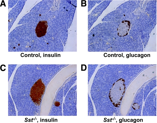
Research design and methods: Islet morphology was assessed by histological analysis. Hormone levels were measured by radioimmunoassay in control and Sst(-/-) mice in vivo and from isolated islets in vitro.

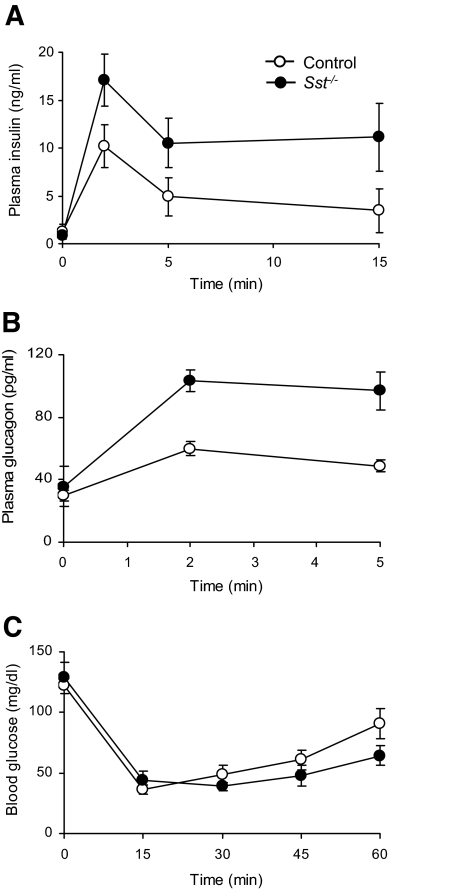
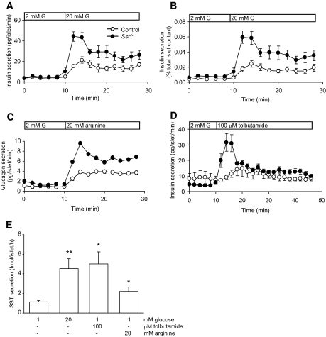
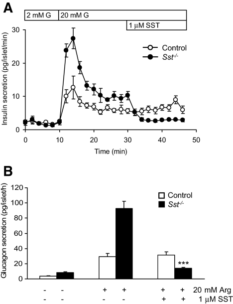
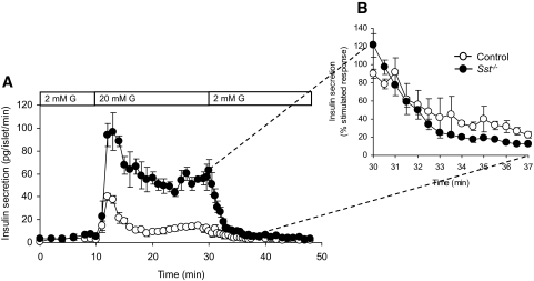
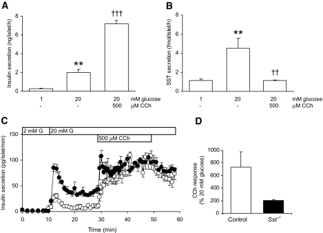
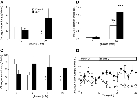
Results: Islet size and organization did not differ between Sst(-/-) and control islets, nor did islet glucagon or insulin content. Sst(-/-) mice showed enhanced insulin and glucagon secretory responses in vivo. In vitro stimulus-induced insulin and glucagon secretion was enhanced from perifused Sst(-/-) islets compared with control islets and was inhibited by exogenous SST in Sst(-/-) but not control islets. No difference in the switch-off rate of glucose-stimulated insulin secretion was observed between genotypes, but the cholinergic agonist carbamylcholine enhanced glucose-induced insulin secretion to a lesser extent in Sst(-/-) islets compared with controls. Glucose suppressed glucagon secretion from control but not Sst(-/-) islets.

Conclusions: We suggest that delta-cell SST exerts a tonic inhibitory influence on insulin and glucagon secretion, which may facilitate the islet response to cholinergic activation. In addition, delta-cell SST is implicated in the nutrient-induced suppression of glucagon secretion.


Figures

FIG. 1.
FIG. 1.
Intraislet organization of α- and β-cells from control and Sst−/− islets. Consecutive sections of control (A and B) or Sst−/− (C and D) mouse islets were stained for insulin (A and C) or glucagon (B and D), respectively, and are representative of sections from three different animals. (Please see http://dx.doi.org/10.2337/db08-0792 for a high-quality digital representation of this figure.)
FIG. 2.
FIG. 2.
Insulin and glucagon secretion in vivo. A: Plasma insulin levels in Sst−/− mice and control mice 0, 2, 5, and 15 min after an intravenous glucose challenge (0.5 g/kg). Points show means ± SE for four to six separate animals. B: Plasma glucagon levels in Sst−/− mice and control mice 0, 2, and 5 min after injection of intravenous arginine (0.25 g/kg). Points show means ± SE for 5 and 14 separate animals (Sst−/− and control, respectively). C: Plasma glucose levels in Sst−/− mice and control mice 0, 15, 30, 45, and 60 min after injection of intravenous insulin (0.4 units/kg). Points show means ± SE for five separate animals.
FIG. 3.
FIG. 3.
Hormone secretion from control and SST−/− islets. A: Insulin secretion from control and Sst−/− mouse islets at a substimulatory concentration of glucose (2 mmol/l G, 0–10 min) and at a stimulatory concentration of glucose (bar, 20 mmol/l G). Points show means ± SE, n = 7–8 separate perifusion channels in each experiment, typical of 10 separate experiments. In B, the same results are expressed as a percentage of total insulin content. C: Arginine-induced (20 mmol/l, bar) glucagon secretion from Sst−/− islets and control islets. Points show means ± SE, n = 4 perifusion channels. Where no error bars are shown, they are smaller than the size of the symbols. D: Insulin secretory responses to the sulfonylurea tolbutamide (100 μmol/l, bar) in the presence of 2 mmol/l glucose from Sst−/− islets and control islets. Points show means ± SE, n = 4 perifusion channels. E: SST secretion from control islets in response to glucose, tolbutamide, and arginine. Bars represent means ± SE, n = 8–9 in one experiment typical of three separate experiments. *P < 0.05, **P < 0.01 vs. 1 mmol/l glucose alone.
FIG. 4.
FIG. 4.
Inhibition of insulin and glucagon secretion by exogenous SST. Effect of exogenous SST (1 μmol/l) on dynamic glucose-induced insulin secretion (20 mmol/l G, bar) (A) and static arginine-induced (20 mmol/l) glucagon secretion (B) from control islets and Sst−/− islets. Points show means ± SE, n = 4 separate perifusion channels in each experiment, typical of six separate experiments. Bars represent means ± SE, n = 6. ***P < 0.001 vs. arginine (Arg) alone.
FIG. 5.
FIG. 5.
Rate of decline in insulin secretion from control and Sst−/− islets after removal of glucose stimulus. A: Glucose-induced (20 mmol/l G, bar) insulin secretion from Sst−/− islets and control islets is reversed to basal levels upon removal of stimulus. B: The decline in insulin secretion from stimulated to basal levels is expressed as percent stimulated insulin secretion before removal of glucose. Points show means ± SE, n = 4 separate perifusion channels.
FIG. 6.
FIG. 6.
Parasympathetic stimulation of insulin secretion from control and Sst−/− islets. Static insulin (A) and SST secretion (B) from control islets in response to glucose (20 mmol/l) and the cholinergic agonist CCh (500 μmol/l). Bars represent means ± SE, n = 8–9 in one experiment typical of three separate experiments. **P < 0.01 vs. 1 mmol/l glucose alone, ††P < 0.01, †††P > 0.001 vs. 20 mmol/l glucose. Data in A and B are from the same experiment. C: Effect of CCh (500 μmol/l) on dynamic glucose-induced (20 mmol/l G, bar) insulin secretion from control (○) and Sst−/− (•) islets. Fractions were collected every 1 min between 10 and 30 min and 50 and 60 min and every 30 s between 30 and 50 min. Points show means ± SE, n = 3 perifusion channels in one experiment representative of three separate experiments. D: The effect of CCh is expressed as a percentage stimulation of the glucose-induced (20 mmol/l) secretory response (mean amplitude of secretion at time points 31–40 min expressed as a percentage of mean amplitude at 21–30 min).
FIG. 7.
FIG. 7.
Glucose-induced suppression of glucagon secretion requires SST. A: Effect of high glucose concentration (20 mmol/l) on glucagon secretion from control islets (□) and Sst−/− islets (▪) in static incubations. Bars represent means ± SE, n = 5–7. *P < 0.05 vs. 2 mmol/l glucose. B: Glucose-induced insulin secretion from the same experiment. **P < 0.01, ***P < 0.001 vs. 2 mmol/l glucose. There was no significant increase in glucagon secretion from Sst−/− islets in response to glucose in seven separate experiments, although in three of these, secretion appeared higher (see A). C: Glucagon secretion from control islets (□) and Sst−/− islets (▪) in response to increasing glucose concentrations. Islets were preincubated in the absence of glucose for 1 h before exposure to the glucose concentrations shown in the figure. Bars represent means ± SE, n = 5–6. *P < 0.05 vs. 0 mmol/l glucose. D: Effect of 10 mmol/l glucose (G, bar) on dynamic glucagon secretion from control islets (○) and Sst−/− islets (•) preincubated in the absence of glucose for 20 min. Points show means ± SE, n = 4 perifusion channels. Controls, P < 0.001 for 0 vs. 10 mmol/l glucose mean values.

Comment in

References

    1. Vieira E, Salehi A, Gylfe E: Glucose inhibits glucagon secretion by a direct effect on mouse pancreatic alpha cells. Diabetologia 50: 370–379, 2007 - PubMed
    1. Gopel SO, Kanno T, Barg S, Rorsman P: Patch-clamp characterisation of somatostatin-secreting δ-cells in intact mouse pancreatic islets. J Physiol 528: 497–507, 2000 - PMC - PubMed
    1. Bishop AE, Polak JM: The anatomy, organization and ultrastructure of the islets of Langerhans. In Textbook of Diabetes. Pickup JC, Williams G, Eds. Oxford, U.K., Blackwell Science, 2002, p. 10.1–10.6
    1. Cabrera O, Berman DM, Kenyon NS, Ricordi C, Berggren PO, Caicedo A: The unique cytoarchitecture of human pancreatic islets has implications for islet cell function. Proc Natl Acad Sci U S A 103: 2334–2339, 2006 - PMC - PubMed
    1. Halban PA, Wollheim CB, Blondel B, Meda P, Niesor EN, Mintz DH: The possible importance of contact between pancreatic islet cells for the control of insulin release. Endocrinology 111: 86–94, 1982 - PubMed

Publication types