. 2008 Oct 31;40(5):550-7.
doi: 10.3858/emm.2008.40.5.550.

Spatiotemporal regulation of fibroblast growth factor signal blocking for endoderm formation in Xenopus laevis

Affiliations

Spatiotemporal regulation of fibroblast growth factor signal blocking for endoderm formation in Xenopus laevis

Sang-wook Cha et al. Exp Mol Med. .

Abstract

We have previously shown that the inhibition of fibroblast growth factor (FGF) signaling induced endodermal gene expression in the animal cap and caused the expansion of the endodermal mass in Xenopus embryos. However, we still do not know whether or not the alteration of FGF signaling controls embryonic cell fate, or when FGF signal blocking is required for endoderm formation in Xenopus. Here, we show that FGF signal blocking in embryonic cells causes their descendants to move into the endodermal region and to express endodermal genes. It is also interesting that blocking FGF signaling between fertilization and embryonic stage 10.5 promotes endoderm formation, but persistent FGF signaling blocking after stage 10.5 restricts endoderm formation and differentiation.


Figures

Figure 1
Figure 1
Effects of blocking FGF signal following DN-FR injection into a single dorsal blastomere in the A, B, C, or D tiers at the 32-cell stage. n-β-galactosidase was used as a tracer. Tadpoles were fixed and stained for β-galactosidase at stage 42. Two representative embryos are shown above. (A) Resultant morphological phenotype was normal when DN-FR was injected into a single blastomere in the A, C, or D tiers. However, embryos injected with DN-FR in the B tier showed a severely malformed phenotype, including a curved back and shortened tail. Interestingly, the data showed the trend that descendants of the DN-FR injected blastomere in A-, C-, and D-tier localized mostly in the endodermal territory. These data are summarized in Table 2. (B) Descendants of a single blastomere in the A tier co-injected with DN-FR and GFP were traced in the endodermal area (Table S1). (C) After DN-FR injection into a single blastomere of each 32-cell stage embryo, their descendants were traced at stage 32 using X-gal staining. Embryos from experiments performed in triplicate were analyzed at stage 32 for the location of n-β-galactosidase positive cells. We counted each region where n-β-galactosidase positive cells were observed densely as 1 but each region where none or several were found as 0 on the sectioned surface of the bisected embryos and the whole embryos. The sum of values of each region in the experimental tadpoles is shown in Table 2. Our analytical method was somewhat subjective, but the results from control embryos agreed well with previously published fate maps (Table 3). (D) The distribution data from Figure 1C were converted into a histogram. The histogram clearly shows that FGF signal deprivation from a single blastomere in the A-tier made most of its descendants move into the endodermal territory instead of their proper location.
Figure 2
Figure 2
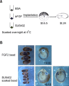
(A) Procedure of the bead implantation assay. (B) Bead implanted embryos were analyzed using whole mount in situ hybridization with Darmin probe, in order to label the mid-gut. An FGF-soaked bead implanted embryo showed the repression of Darmin expression around the bead (15/16 affected embryos/total embryos). However, cells neighboring the SU5402 soaked bead showed strong expression of darmin (18/18 affected embryos/total embryos). This shows that the presence or absence of FGF signaling is strongly related to the expression of endodermal genes in embryonic cells during Xenopus embryogenesis (Table S2).
Figure 3
Figure 3
(A) Schematic diagram of the time frame of SU5402 treatment of Xenopus embryos. (B) Morphology of embryos treated with SU5402 according to the designed time frame. Most of the embryos that were treated with SU5402 before stage 10.5 (groups A, B, and C) showed an expanded abdominal region. However, a curved back and shortened tail were seen in the group treated with SU5402 after stage 10.5 (group E). (C) Gene expression pattern of embryos treated with SU5402 according to the time frame. Expression of endodermin appeared to be normal in the case of FGF signal deprivation in embryos from fertilization to stage 10.5. However, its expression was abruptly decreased when the FGF signal was blocked after stage 10.5. This shows that FGF signal blockade before stage 10.5 is critically important for proper endoderm formation but persistent blocking of FGF signal after stage 10.5 inhibited proper endoderm formation. When the FGF signaling was blocked after stage 10.5, neural induction was inhibited as shown (Group D, E, F), and the expression of actin, a general mesodermal marker, was also reduced (group E, F). (D) The future endodermal region of the vegetal hemispheres was dissected from embryos at MBT and conjugated with animal cap explants. The conjugated explants were cultured with FGF signal blocking until stage 30. As shown in Group A, the expression of the general endodermal marker endodermin was increased by RT-PCR analysis but the expression of endodermal organ specific markers such as Xlhbox8 and IFABP were not observed when compared with conjugated explants cultured without SU5402. The RT-PCR results showed that the explants remained in the initial stage of the endodermalization instead of differentiating into the specific organ cells as shown at Group B.

Similar articles

Cited by

References

    1. Agius E, Oelgeschlager M, Wessely O, Kemp C, De Robertis EM. Endodermal Nodal-related signals and mesoderm induction in Xenopus. Development. 2000;127:1173–1183. - PMC - PubMed
    1. Amaya E, Stein PA, Musci TJ, Kirschner MW. FGF signalling in the early specification of mesoderm in Xenopus. Development. 1993;118:477–487. - PubMed
    1. Cha SW, Hwang YS, Chae JP, Lee SY, Lee HS, Daar I, Park MJ, Kim J. Inhibition of FGF signaling causes expansion of the endoderm in Xenopus. Biochem Biophys Res Commun. 2004;315:100–106. - PubMed
    1. Ciruna B, Rossant J. FGF signaling regulates mesoderm cell fate specification and morphogenetic movement at the primitive streak. Dev Cell. 2001;1:37–49. - PubMed
    1. Clements D, Friday RV, Woodland HR. Mode of action of VegT in mesoderm and endoderm formation. Development. 1999;126:4903–4911. - PubMed

Publication types

MeSH terms

LinkOut - more resources