

Amygdala-frontal connectivity during emotion regulation
- PMID: 18985136
- PMCID: PMC2566753
- DOI: 10.1093/scan/nsm029
Amygdala-frontal connectivity during emotion regulation
Abstract
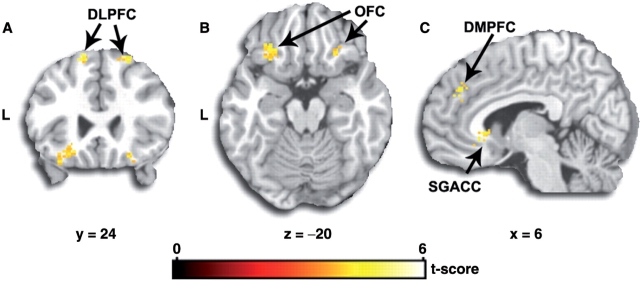
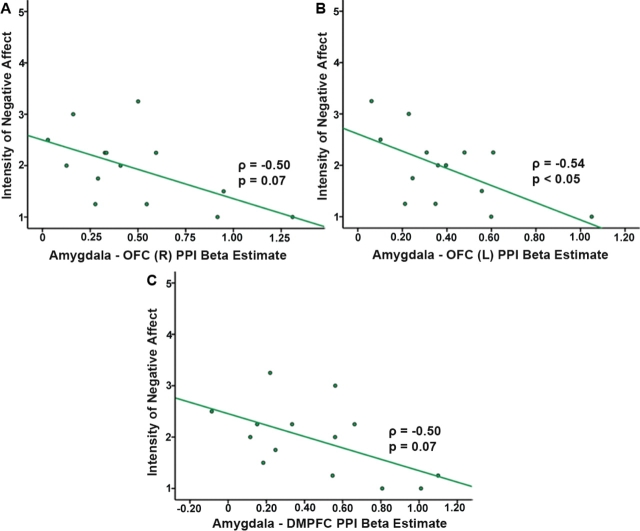
Successful control of affect partly depends on the capacity to modulate negative emotional responses through the use of cognitive strategies (i.e., reappraisal). Recent studies suggest the involvement of frontal cortical regions in the modulation of amygdala reactivity and the mediation of effective emotion regulation. However, within-subject inter-regional connectivity between amygdala and prefrontal cortex in the context of affect regulation is unknown. Here, using psychophysiological interaction analyses of functional magnetic resonance imaging data, we show that activity in specific areas of the frontal cortex (dorsolateral, dorsal medial, anterior cingulate, orbital) covaries with amygdala activity and that this functional connectivity is dependent on the reappraisal task. Moreover, strength of amygdala coupling with orbitofrontal cortex and dorsal medial prefrontal cortex predicts the extent of attenuation of negative affect following reappraisal. These findings highlight the importance of functional connectivity within limbic-frontal circuitry during emotion regulation.
Figures
References
-
- Adolphs R. Neural systems for recognizing emotion. Current Opinion in Neurobiology. 2002;12:169–77. - PubMed
-
- Aggleton JP. The contribution of the amygdala to normal and abnormal emotional states. Trends in Neuroscience. 1993;16:328–33. - PubMed
-
- Amaral DG, Bauman MD, Capitanio JP, et al. The amygdala: is it an essential component of the neural network for social cognition? Neuropsychologia. 2003;41:517–22. - PubMed
-
- Amaral DG, Price JL. Amygdalo-cortical projections in the monkey (Macaca fascicularis) Journal of Comparative Neurology. 1984;230:465–96. - PubMed
-
- Angrilli A, Mauri A, Palomba D, et al. Startle reflex and emotion modulation impairment after a right amygdala lesion. Brain. 1996;119:1991–2000. - PubMed
Publication types
MeSH terms
Grants and funding
LinkOut - more resources
Full Text Sources
Other Literature Sources
Medical
