

Distinct regions of medial rostral prefrontal cortex supporting social and nonsocial functions
- PMID: 18985143
- PMCID: PMC2569804
- DOI: 10.1093/scan/nsm014
Distinct regions of medial rostral prefrontal cortex supporting social and nonsocial functions
Abstract
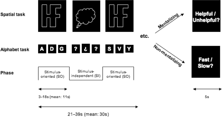
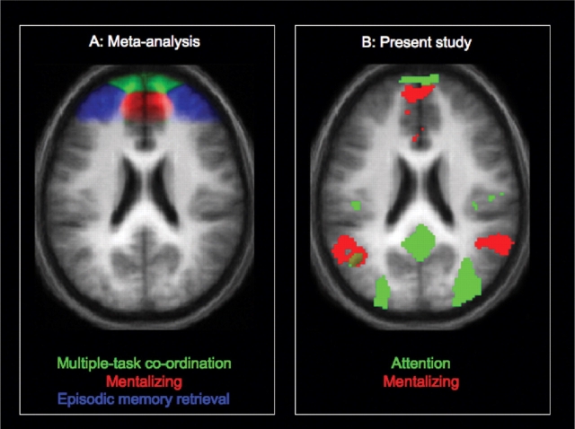
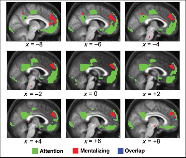
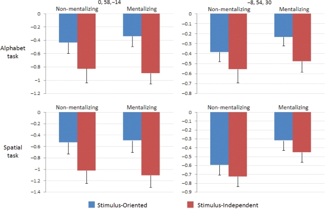
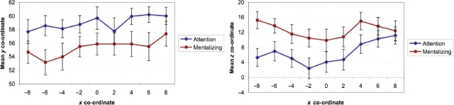
While some recent neuroimaging studies have implicated medial rostral prefrontal cortex (MPFC) in 'mentalizing' and self-reflection, others have implicated this region in attention towards perceptual vs self-generated information. In order to reconcile these seemingly contradictory findings, we used fMRI to investigate MPFC activity related to these two functions in a factorial design. Participants performed two separate tasks, each of which alternated between 'stimulus-oriented phases' (SO), where participants attended to task-relevant perceptual information, and 'stimulus-independent phases' (SI), where participants performed the same tasks in the absence of such information. In half of the blocks ('mentalizing condition'), participants were instructed that they were performing these tasks in collaboration with an experimenter; in other blocks ('non-mentalizing condition'), participants were instructed that the experimenter was not involved. In fact, the tasks were identical in these conditions. Neuroimaging data revealed adjacent but clearly distinct regions of activation within MPFC related to (i) mentalizing vs non-mentalizing conditions (relatively caudal/superior) and (ii) SO vs SI attention (relatively rostral/inferior). These results generalized from one task to the other, suggesting a new axis of functional organization within MPFC.
Figures
References
-
- Amodio DM, Frith CD. Meeting of minds: the medial frontal cortex and social cognition. Nature Reviews Neuroscience. 2006;7:268–77. - PubMed
-
- Barbas H, Ghashghaei H, Dombrowski SM, Rempel-Clower NL. Medial prefrontal cortices are unified by common connections with superior temporal cortices and distinguished by input from memory-related areas in the rhesus monkey. Journal of Comparative Neurology. 1999;410:343–67. - PubMed
-
- Bird CM, Castelli F, Malik O, Frith U, Husain M. The impact of extensive medial frontal lobe damage on ‘theory of mind’ and cognition. Brain. 2004;127:914–28. - PubMed
-
- Burgess PW. Strategy application disorder: the role of the frontal lobes in human multitasking. Psychological Research. 2000;63:279–88. - PubMed
-
- Burgess PW, Dumontheil I, Gilbert SJ, Okuda J, Schölvinck ML, Simons JS. On the role of rostral prefrontal cortex (area 10) in prospective memory. In: Kliegel M, McDaniel MA, Einstein GO, editors. Prospective Memory: Cognitive, Neuroscience, Developmental, and Applied Perspectives. Mahwah: Erlbaum; in press.
Publication types
MeSH terms
Grants and funding
LinkOut - more resources
Full Text Sources
