. 2007 Sep;2(3):240-50.
doi: 10.1093/scan/nsm020.

The right-hemisphere and valence hypotheses: could they both be right (and sometimes left)?

Affiliations

The right-hemisphere and valence hypotheses: could they both be right (and sometimes left)?

William D S Killgore et al. Soc Cogn Affect Neurosci. 2007 Sep.

Abstract

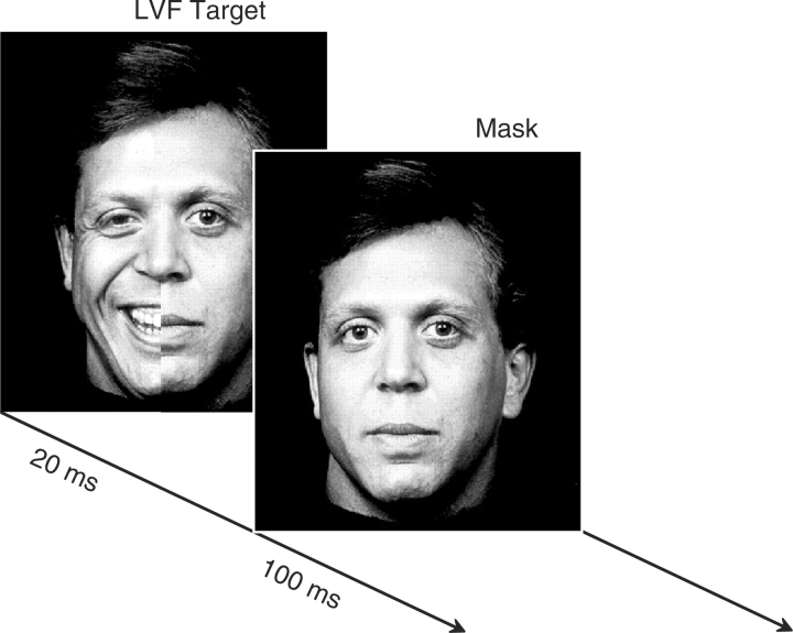
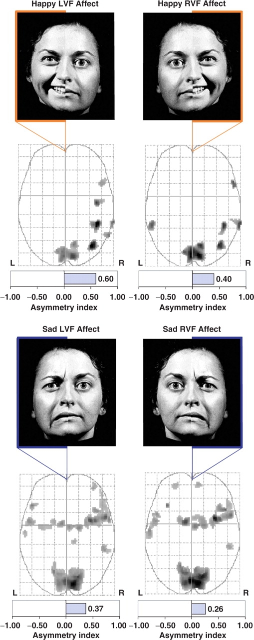
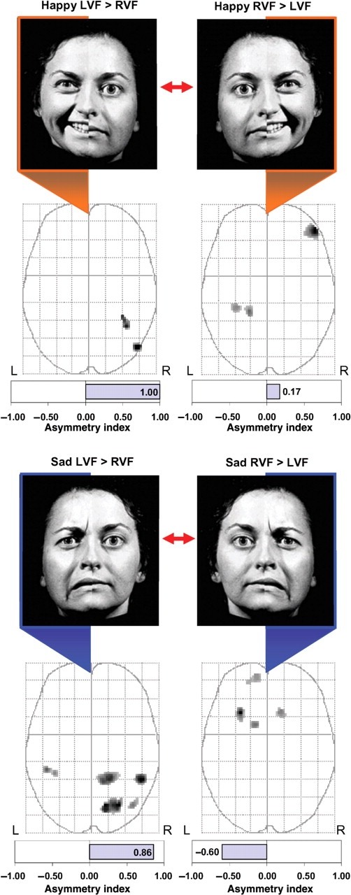
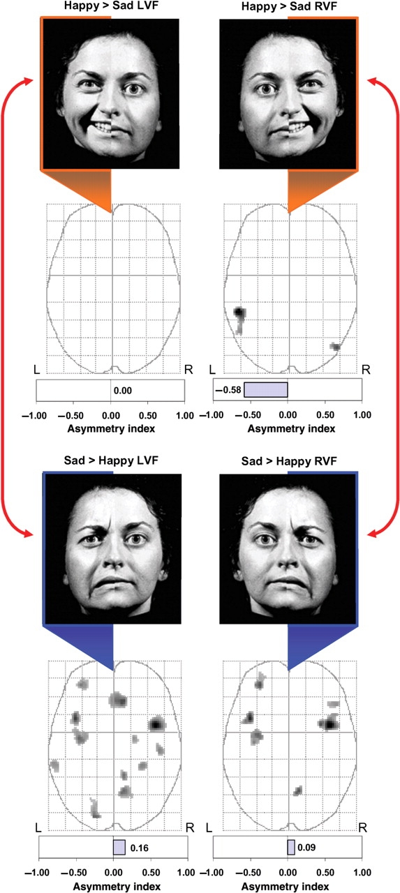
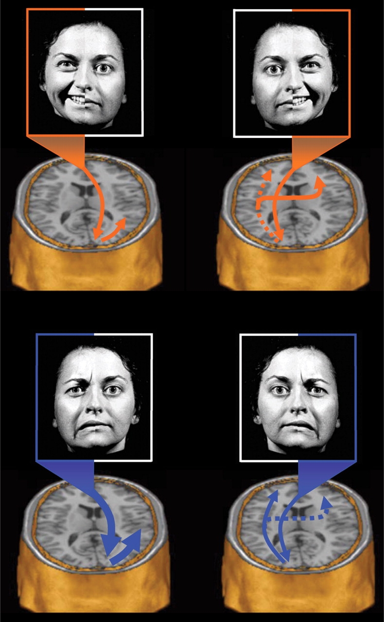
The two halves of the brain are believed to play different roles in emotional processing, but the specific contribution of each hemisphere continues to be debated. The right-hemisphere hypothesis suggests that the right cerebrum is dominant for processing all emotions regardless of affective valence, whereas the valence specific hypothesis posits that the left hemisphere is specialized for processing positive affect while the right hemisphere is specialized for negative affect. Here, healthy participants viewed two split visual-field facial affect perception tasks during functional magnetic resonance imaging, one presenting chimeric happy faces (i.e. half happy/half neutral) and the other presenting identical sad chimera (i.e. half sad/half neutral), each masked immediately by a neutral face. Results suggest that the posterior right hemisphere is generically activated during non-conscious emotional face perception regardless of affective valence, although greater activation is produced by negative facial cues. The posterior left hemisphere was generally less activated by emotional faces, but also appeared to recruit bilateral anterior brain regions in a valence-specific manner. Findings suggest simultaneous operation of aspects of both hypotheses, suggesting that these two rival theories may not actually be in opposition, but may instead reflect different facets of a complex distributed emotion processing system.


Figures

Fig. 1
Fig. 1
Masked chimeric face stimuli. Each trial consisted of two stimuli presented in rapid succession: (i) a ‘target’ chimeric face depicting an emotional expression on one half and a neutral expression from the same poser on the other half. There was an equal number of presentations of left-sided and right-sided stimuli. Each target chimeric expression was presented for 20 ms and was immediately replaced by (ii) a ‘mask’ face consisting of a photograph of the same poser expressing a neutral emotion for 100 ms. Participants provided feedback on each trial as to whether the poser was a male or female. Each trial was separated by a 3 s interstimulus interval.
Fig. 2
Fig. 2
One sample t-tests for chimeric stimuli. Brain activity associated with each unilateral affective stimulation condition was tested relative to resting baseline using a one sample t-test (P < 0.001, uncorrected). The top row shows happy chimera and the bottom row shows sad chimera. Relative to resting baseline, brain activity was greater in the right hemisphere during all four affective hemiface conditions regardless of affective valence (i.e. happy vs sad) or visual field of presentation [i.e. Left Visual Field (LVF) vs Right Visual Field (RVF)]. Maximum intensity projections (MIPs) in the axial plane show similar activity in the primary visual cortex and greater right hemisphere activity for all four conditions.
Fig. 3
Fig. 3
Comparisons between lateralized stimulus conditions. Brain activity associated with unilateral LVF presentations was compared directly to activity associated with unilateral RVF presentations using paired t-tests. Regions showing significant differences (P < 0.005, uncorrected) between the two lateralized presentation conditions are displayed on the MIPs in the axial plane. Happy hemifaces presented to the LVF produced significantly greater activity in the posterior right hemisphere (fusiform gyrus and middle temporal gyrus) than when presented to the RVF (top left). In contrast, happy hemifaces presented to the RVF produced greater activity in the right lateral orbitofrontal cortex and left fusiform/parahippocampal gyrus (top right). For sad hemifaces, LVF presentations produced greater activity predominantly in the posterior right hemisphere compared to similar RVF presentations (bottom left). In contrast, RVF presentations produced greater activity in the left orbitofrontal cortex and basal ganglia when compared to identical LVF presentations (bottom right).
Fig. 4
Fig. 4
Comparisons between valence conditions. Brain activity associated with happy vs sad stimuli restricted to a single visual field was compared using paired t-tests (P < 0.005, uncorrected) and displayed on MIPs in the axial plane. For LVF stimuli, there were no regions where unilateral happy hemifaces produced greater activation than unilateral sad hemifaces. In contrast, sad hemifaces presented to the LVF produced significantly greater activity across a distributed bilateral network of affective brain regions when compared activity produced by matched happy hemifaces. For RVF stimuli, happy hemifaces produced significantly greater activity in bilateral middle temporal gyri, whereas sad hemifaces restricted to the RVF produced significantly greater activation within several distributed affect regions of both hemispheres when compared to comparable happy hemifaces.
Fig. 5
Fig. 5
Proposed model of regional interactions. The present findings were used to outline a tentative model whereby the posterior right hemisphere is dominant for processing all facial affective stimuli regardless of valence, but is also particularly specialized for processing negative affective stimuli. In contrast, the posterior left hemisphere is postulated to be relatively less effective at processing affective stimuli in general and must, therefore, rely on downstream processing within the prefrontal cortex bilaterally to evaluate the significance of the affective stimulus. This prefrontal system appears to recruit the left and right anterior regions in a valence specific manner. Top left: Happy affective stimuli in the LVF are initially projected to the right hemisphere primary visual cortex and then intra-hemispherically to nearby posterior temporal and fusiform regions for further analysis. Top right: In contrast, happy stimuli in the RVF are first projected to the primary visual cortex of the left hemisphere. Because the left hemisphere is relatively less specialized for processing facial affect, such information is sent downstream for further elaborative processing. Happy affect in this hemisphere appears to activate left fusiform and left middle temporal gyri and is further projected bilaterally to the prefrontal cortices for elaboration and comparison, leading to valence specific activation of the right prefrontal cortex. Bottom left: Sad affective stimuli from the LVF would be directly projected to the primary visual cortex of the right hemisphere. Because of the superiority of the right hemisphere for processing affect, and negative affect in particular, very little transfer distance would be required for valence-specific elaboration. Consequently, negative facial affect cues in the LVF would be expected to be more rapidly and efficiently processed than any other affect/visual field combination. Bottom right: In contrast, sad affective stimuli in the RVF would be particularly disadvantaged, as they would be sent to the non-affect dominant left hemisphere. Due to the relative non-specialization of the left hemisphere for processing affect, the information would be subsequently relayed to the anterior regions for further elaboration and comparison. Because there exist many more categories of negative emotion than positive, the processing of negative affect in the left hemisphere is likely to be particularly inefficient. Globally, this model predicts that LVF presentations should generally be superior to RVF presentations, regardless of valence, but further suggests that RVF presentations of happy expressions will typically result in superior processing than identical presentations of sad expressions.

Similar articles

Cited by

References

    1. Adolphs R, Damasio H, Tranel D, Cooper G, Damasio AR. A role for somatosensory cortices in the visual recognition of emotion as revealed by three-dimensional lesion mapping. Journal of Neuroscience. 2000;20:2683–90. - PMC - PubMed
    1. Adolphs R, Damasio H, Tranel D, Damasio AR. Cortical systems for the recognition of emotion in facial expressions. Journal of Neuroscience. 1996;16:7678–87. - PMC - PubMed
    1. Adolphs R, Jansari A, Tranel D. Hemispheric perception of emotional valence from facial expressions. Neuropsychology. 2001;15:516–24. - PubMed
    1. Ahern GL, Schwartz GE. Differential lateralization for positive vs negative emotion. Neuropsychologia. 1979;17:693–8. - PubMed
    1. Borod JC, Cicero BA, Obler LK, et al. Right hemisphere emotional perception: evidence across multiple channels. Neuropsychology. 1998;12:446–58. - PubMed

Publication types