. 2008;3(11):e3632.
doi: 10.1371/journal.pone.0003632. Epub 2008 Nov 5.

Retinoblastoma loss modulates DNA damage response favoring tumor progression

Affiliations

Retinoblastoma loss modulates DNA damage response favoring tumor progression

Marcos Seoane et al. PLoS One. 2008.

Abstract

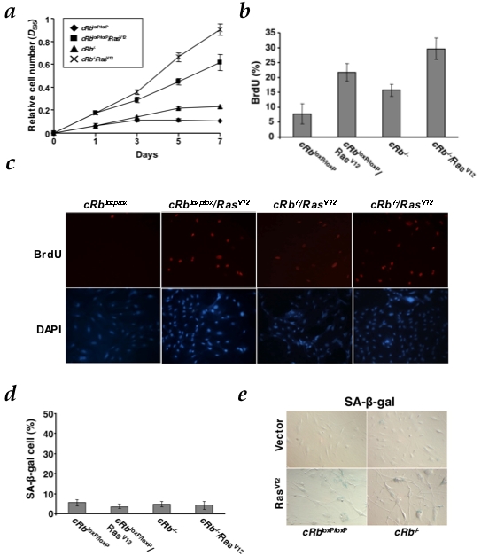
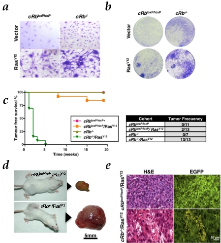
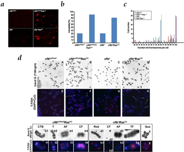
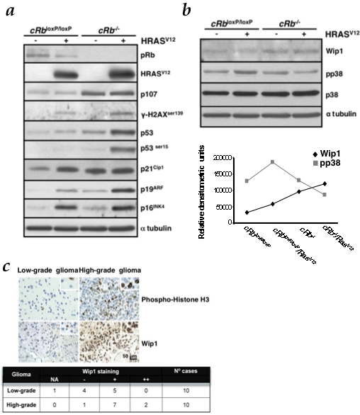
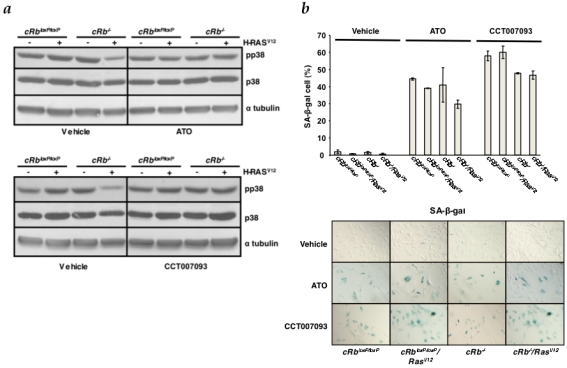
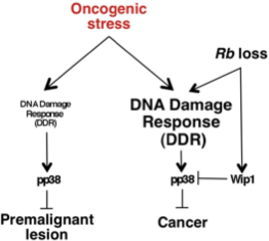
Senescence is one of the main barriers against tumor progression. Oncogenic signals in primary cells result in oncogene-induced senescence (OIS), crucial for protection against cancer development. It has been described in premalignant lesions that OIS requires DNA damage response (DDR) activation, safeguard of the integrity of the genome. Here we demonstrate how the cellular mechanisms involved in oncogenic transformation in a model of glioma uncouple OIS and DDR. We use this tumor type as a paradigm of oncogenic transformation. In human gliomas most of the genetic alterations that have been previously identified result in abnormal activation of cell growth signaling pathways and deregulation of cell cycle, features recapitulated in our model by oncogenic Ras expression and retinoblastoma (Rb) inactivation respectively. In this scenario, the absence of pRb confers a proliferative advantage and activates DDR to a greater extent in a DNA lesion-independent fashion than cells that express only HRas(V12). Moreover, Rb loss inactivates the stress kinase DDR-associated p38MAPK by specific Wip1-dependent dephosphorylation. Thus, Rb loss acts as a switch mediating the transition between premalignant lesions and cancer through DDR modulation. These findings may have important implications for the understanding the biology of gliomas and anticipate a new target, Wip1 phosphatase, for novel therapeutic strategies.


Conflict of interest statement

Competing Interests: The authors have declared that no competing interests exist.

Figures

Figure 1
Figure 1. Mouse astrocytes are defective for HRasv12-induced cell cycle arrest.
a, Growth curve analysis of early-passage cRb loxP/loxP conditional astrocytes co-infected with PIG/pBABE (vector), PIG/pBABE-HRasV12, PIG-Cre/pBABE and PIG-Cre/pBABE-HRasV12 retroviral vectors. After infection, cells were plated in triplicate and the cells were fixed on the indicated days for subsequent staining with crystal violet. Each time point represents the mean±s.d. of total cumulative cell number from at least three independent experiments. b, Measurement of the proliferation of astrocytes by BrdU incorporation assay (panel c). c, Cells were labeled with BrdU for 5 h on day 5 after puromycin selection. d, Senescence assays in cRbloxP/loxP and cRb−/−. The y-axis represents the percentage of SA-β-galactosidase-positive cells (mean and s.d.) from at least three independent experiments. e, A representative result of three independent experiments is shown. Photographs are at the same magnification.
Figure 2
Figure 2. HRasV12 in the absence of Rb induces malignant transformation both in vitro and in vivo.
a, Ability to form foci, representative pictures of wells stained with crystal violet (day 7). b, Representative pictures of wells stained on the indicated days of culture showing the morphological change observed in each group are also shown. c, Tumor-free survival curve (Kaplan-Meier plot) for tumor formation in SCID mice. Tumor was found in cRb −/−/RasV12 cohort (P<0.0001) d, Photographs of SCID mice after subcutaneous injection of cRb loxP/loxP/RasV12 and cRb −/−/RasV12 astrocytes at eleven and two weeks respectively. Actual sizes of tumors after biopsy. e. Low-grade gliomas (cRb loxP/loxP/RasV12; H&E, upper left) showed a rather monomorphic appearance, with polygonal or rounded cell shape and smaller rounded nuclei without prominent nucleoli. Note the absence of mitosis and necrosis. In contrast, high-grade gliomas (cRb−/−/RasV12; H&E, lower left) displayed cellular pleomorphism, with large, fusiform and irregular nuclei that frequently exhibit mitotic figures (upper left corner) and necrotic foci (lower right corner). Co-expression of EGFP permits the visualization of the previously modified astrocytes.
Figure 3
Figure 3. HRasV12-dependent production of reactive oxygen species (ROS) and associated chromosomal instability.
a, cells were assayed for dihydroethidium (DHE) fluorescence, indicative of ROS production and visualized by fluorescent microscopy. Under identical imaging conditions, DHE oxidation was significantly increased in cRbloxP/loxP/RasV12 and cRb−/−/RasV12 astrocytes. b, cRb−/−/RasV12 and cRbloxP/loxP/RasV12 cells are significantly more aneuploid than cRb loxP/loxP and cRb −/− cells. c, Absolute chromosome numbers of cRb loxP/loxP, cRb loxP/loxP/RasV12, cRb −/− and cRb −/−/RasV12 cells, showing the presence of diploid and tetraploid populations. Representative data of at least three independent experiments. d, Band G (Wright S) and telomere fluorescence in situ hybridization (T-Fish) was performed using a Cy3-labeled peptide nucleic acid (PNA) telomeric probe. Representative metaphase spreads from cRb loxP/loxP and cRb −/−, partial representative metaphase spreads from cRb loxP/loxP/RasV12 and cRb−/−/RasV12 stained with Wright and hybridized with Cy3 telomeric probe are shown. The regions of metaphase spreads from cRb loxP/loxP/RasV12 and cRb −/−/RasV12 are magnified in the lower panel to show centromere fragments (CF), acentric fragment (AF), chromatide type break (CTB), telomeric fusion (TF), robertsonian translocation (Rob) and other type of translocation (T). X indicates quadrivalent (Qua) found in cRb loxP/loxP/RasV12.
Figure 4
Figure 4. Rb loss enhances DNA damage response (DDR) induced by oncogenic Ras and reduces the p-p38MAPK levels by upregulation of Wip1.
a, HRasV12 is able to induce DDR markers expression, such as p16INK4a, p21Cip1 and p19ARF, p53 and p53ser15 and γ-H2AXser139. Rb loss, in the presence of oncogenic Ras, increases this response. Immunoblot analysis was performed on astrocytes lysates prepared at day 6 after co-infection and selection with puromycin. b, Immunoblot analysis of p-p38MAPK and Wip1 levels was performed on cell lysates prepared at day 5 from co-infection and selection with puromycin. Densitometric analysis (in relative densitometric units) of Wip1 and p-p38MAPK protein levels. c, TMA analysis of Wip1 expression in glioma clinical samples. Representative pictures of samples are shown. The table shows median values. Wip1 immunoreactivity intensity was assigned according to the following scale: NA, non-assessable cases; −, less than 10% of neoplastic cells displayed immunoreactivity; +, 11–29% of neoplastic cells displayed immunoreactivity; ++, 30% or greater percentage of neoplastic cells displayed immunoreactivity. Phospho-histone H3 was used as a proliferation marker. Scale bars, 50 µm.
Figure 5
Figure 5. Wip1 inhibition enables senescence in cRb−/−/RasV12 astrocytes.
a, All the experimental groups were treated with the Wip1 chemical inhibitors CCT007093 and Arsenic Trioxide (ATO). Levels of p-p38MAPK were assessed by Western blot analysis as a readout of Wip1 phosphatase activity inhibition. b, SA-β-galactosidase assays in all groups (without treatment, ATO treatment, CCT007093 treatment). The y-axis represents the percentage of-positive cells (mean and s.d.).
Figure 6
Figure 6. DDR is modulated by Rb loss switching premalignant lesion to cancer.

Similar articles

Cited by

References

    1. Kleihues P, Burger PC, Scheithauer BW. The new WHO classification of brain tumours. Brain Pathol. 1993;3(3):255–268. - PubMed
    1. Guha A, Feldkamp MM, Lau N, Boss G, Pawson A. Proliferation of human malignant astrocytomas is dependent on ras activation. Oncogene. 1997;15(23):2755–2765. - PubMed
    1. Feldkamp MM, Lau N, Rak J, Kerbel RS, Guha A. Normoxic and hypoxic regulation of vascular endothelial growth factor (VEGF) by astrocytoma cells is mediated by ras. Int J Cancer. 1999;81(1):118–124. - PubMed
    1. Rajasekhar VK, Viale A, Socci ND, Wiedmann M, Hu X, et al. Oncogenic ras and akt signaling contribute to glioblastoma formation by differential recruitment of existing mRNAs to polysomes. Mol Cell. 2003;12(4):889–901. - PubMed
    1. Hu X, Pandolfi PP, Li Y, Koutcher JA, Rosenblum M, et al. mTOR promotes survival and astrocytic characteristics induced by Pten/AKT signaling in glioblastoma. Neoplasia. 2005;7(4):356–368. - PMC - PubMed

Publication types

MeSH terms

Substances