Case Reports
. 2008;3(11):e3642.
doi: 10.1371/journal.pone.0003642. Epub 2008 Nov 5.

The zinc transporter SLC39A13/ZIP13 is required for connective tissue development; its involvement in BMP/TGF-beta signaling pathways

Affiliations
Case Reports

The zinc transporter SLC39A13/ZIP13 is required for connective tissue development; its involvement in BMP/TGF-beta signaling pathways

Toshiyuki Fukada et al. PLoS One. 2008.

Erratum in

Abstract

Background: Zinc (Zn) is an essential trace element and it is abundant in connective tissues, however biological roles of Zn and its transporters in those tissues and cells remain unknown.

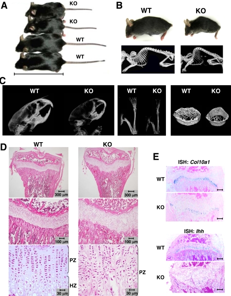
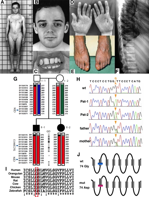
Methodology/principal findings: Here we report that mice deficient in Zn transporter Slc39a13/Zip13 show changes in bone, teeth and connective tissue reminiscent of the clinical spectrum of human Ehlers-Danlos syndrome (EDS). The Slc39a13 knockout (Slc39a13-KO) mice show defects in the maturation of osteoblasts, chondrocytes, odontoblasts, and fibroblasts. In the corresponding tissues and cells, impairment in bone morphogenic protein (BMP) and TGF-beta signaling were observed. Homozygosity for a SLC39A13 loss of function mutation was detected in sibs affected by a unique variant of EDS that recapitulates the phenotype observed in Slc39a13-KO mice.

Conclusions/significance: Hence, our results reveal a crucial role of SLC39A13/ZIP13 in connective tissue development at least in part due to its involvement in the BMP/TGF-beta signaling pathways. The Slc39a13-KO mouse represents a novel animal model linking zinc metabolism, BMP/TGF-beta signaling and connective tissue dysfunction.


Conflict of interest statement

Competing Interests: The authors have declared that no competing interests exist.

Figures

Figure 1
Figure 1. Growth retardation, kyphosis, osteopenia, and abnormal cartilage development in Slc39a13-KO mice.
A. 5-week-old female wild-type and Slc39a13-KO mice. A bar indicates 10 cm. B. Kyphosis in Slc39a13-KO mouse. Appearance and radiographs of 5-week-old mice. C. Osteopenia of Slc39a13-KO mice. X-rays of skull (left), femur and tibia (middle) of 4-week-old mice. 3D-mCT of the tibial diaphysis (right). D. Slc39a13-KO mice show elongated growth plate with uncoordinated columnar formation, and decrease hypertrophic zone. Tibia from 4-week-old Slc39a13-KO mice and wild-type littermates stained with H&E. Large views of growth plate are shown in middle and lower panels. PZ, proliferative zone; HZ, hypertrophic zone. E. Gene expression of Col10a1 and Ihh gene are diminished in growth plate of 4-week-old Slc39a13-KO mice. Bar indicates 300 µm. ISH images.
Figure 2
Figure 2. Abnormal teeth and craniofacial development in Slc39a13-KO mice.
A. Slc39a13-KO mice develop abnormal incisor teeth (upper). 5-week-old Slc39a13-KO mice show evidence of malocclusion (red arrow head), deformity (blue arrow head), and breakage (yellow arrow head) of incisor teeth. Lower panels show 3D m-CT imaging analysis showing a systemic decrease in bone density and abnormal tooth development of 5-week-old Slc39a13-KO mice compared with wild-type littermates. B. Craniofacial features of Slc39a13-KO mouse. Eye shows enophthalmos-like appearance and downslanting palpebral fissures in Slc39a13-KO mouse. Representative face images of 5-week-old wild-type and Slc39a13-KO mouse. C. Root dentin formation of molar teeth (red arrow head) and the bone volume fraction of mandible (yellow arrow head) are remarkably reduced in Slc39a13-KO mice. BEN (upper) and 3D m-CT images (lower) of mandibular molar regions in 5-week-old wild-type and Slc39a13-KO mice are shown. D. Root dentin and alveolar bone are reduced in Slc39a13-KO mice. Sagittal sections of mandibular molar regions in 5-week-old wild-type and Slc39a13-KO mice were stained with H&E. Boxed areas are reproduced at higher magnification. de: dentin, ab: alveolar bone.
Figure 3
Figure 3. Decreased dermal and corneal stromal collagen in Slc39a13-KO mice.
A. Dermal collagen is decreased in Slc39a13-KO mice. H&E staining shows 5-week-old Slc39a13-KO skin is thinner than wild-type littermates (left), without significant difference in epidermis (right; enlarged view of blue-boxed area 1). Azan staining shows collagen fibril is decreased in Slc39a13-KO skin (middle). B. Fragility of skin is increased in Slc39a13-KO mice. The strength of skin under tension is weakened in 5-week-old Slc39a13-KO mice compared with wild-type littermates (n = 3 for each). Data represent mean±S.D. C. Magnified image of green-boxed area 2 in Figure 3A shows morphology of dermal fibroblasts (red arrow) are spindle-shaped and often stellate with cytoplasmic extension in wild-type, while they are mostly round to oval in Slc39a13-KO mice. D. TEM images of transversely sectioned dermal collagen from 5-week-old mice are shown. Bar; 500 nm. E. Dermal collagen in 5-week-old Slc39a13-KO mice are characterized by thinner in size (left), and lower density of collagen fibrils (right) than that in a wild-type mice. Thirty-nine and 168 areas in TEM images of wild-type and Slc39a13-KO samples, respectively were assessed. Data represent mean±S.D. F. Collagen of corneal stroma is decreased in Slc39a13-KO mice. H&E (left) and Azan (middle) staining show 5-week-old Slc39a13-KO cornea is thinner than wild-type littermates. Magnified images of red-boxed area in middle panel show the width of corneal stroma (substantia propia; SP) is decreased in Slc39a13-KO cornea, without significant difference in corneal epithelial cells (CEP) (right). G. Relative ratio between SP versus CEP indicates significant reduction of stromal collagen in Slc39a13-KO cornea compared with those of wild-type (n = 5 for each). Data represent mean±S.D.
Figure 4
Figure 4. Clinical features, and genetic and molecular evidence of the SLC39A13 mutation in the two subjects with short stature and EDS.
The elder affected sib is shown at age 22 with short stature and with mildly shortened trunk (A), antimongoloid eye slant with lack of periorbital tissue (B), missing upper lateral incisor tooth (C), thin and finely wrinkled skin on the palm of his hands (D), severe varicosity of his lower legs and feet (E), vertebral flattening with sclerosis of the vertebral endplates (F, radiograph taken at age 18 years). G. Autozygous haplotype blocks detected by SNP genotyping are represented by the black boxes, while blocks detected by microsatellite analyses are represented as solid boxes in different colors (the common ancestral haplotype being represented in red). The microsatellites used are shown on the left, and the numbers refer to microsatellites' alleles. Gene density (GD) for local transcripts is shown in the center. Individual genes are depicted as small circles, shaded (for the olfactory receptor genes clusters) or open (all other genes). The relative position of SLC39A13 and of the centromere (CEN) are also indicated. H. Sequence tracings from SLC39A13 amplicons showing homozygosity for a G to A transition changing codon 74 from glycine to aspartic acid in the two affected subjects (Pat-1 and Pat-2), as well as heterozygosity in the parents. I. Alignment of amino acid sequences of SLC39A13 protein showing high conservation of sequences and in particular of glycine-74 (red box). J. Assignment of transmembrane domains of SLC39A13 (shaded boxes) was taken from Uniprot (http://www.uniprot.org). The substitution of glycine-74 with aspartic acid was predicted to cause a six-residue shift of the second transmembrane domain towards the carboxy end by HMMTOP program (http://www.enzim.hu/hmmtop/, lower panel).
Figure 5
Figure 5. Cellular localization of Slc39a13.
A. Slc39a13 gene is expressed in osteoblasts (1 and 2; red arrow head) in tibia of 4-week-old mice. Regions indicated as 1 and 2 in top are magnified as 400 times. ISH images. B. Slc39a13 mRNA is expressed in proliferative zone of growth plate (left) at 3-week-old tibia. Region indicated as 1 and 2 in top are enlarged at lower panels. PZ: proliferative zone. HZ: hypertrophic zone. C. Slc39a13 gene is expressed in odontoblasts of 10-day-old molar teeth. ISH analysis shows Slc39a13 is expressed in odontoblast (black arrowhead) lining the dentin of crown (1), and in osteoblast (red arrowhead) on the surface of alveolar bone (2). Regions indicated as 1 and 2 in top are magnified as 400 times. od: odontoblasts, ab: alveolar bone. D. Slc39a13 protein expression in dermal fibroblasts. IHC analysis shows Slc39a13 protein is expressed in fibroblasts in dermis of 5-week-old wild-type mice (left upper). Enlarged images of boxed areas are shown in IHC images. Bar indicates 10 µm.
Figure 6
Figure 6. Slc39a13 controls intracellular zinc distribution.
A. Slc39a13 protein locates on perinuclear region in primary osteoblasts (1), chondrocytes (2), pulpal cells (3), and dermal fibroblasts (4). Slc39a13, nuclei, and actin were stained with anti-Slc39a13 specific antibody, DAPI, and phalloidin, respectively. Confocal microscopic images. B. Slc39a13 is localized in Golgi. Confocal microscopic analysis using dermal fibroblasts. Golgi, Slc39a13, nuclei, and actin is stained with anti-GM130 antibody, anti-Slc39a13 specific antibody, DAPI, and phalloidin, respectively. C. Upper: Schematic diagram of EPMA to detect intracellular zinc level. Golgi and nucleus in primary dermal fibroblasts are scanned by electron beam (EB), and characteristic X-ray (CX) for zinc is detected. Lower: Slc39a13 involves in control of intracellular zinc distribution. The ratio of zinc level in Golgi or nucleus versus total Zn in those organelles (n = 11 and 10 for wild-type and Slc39a13-KO dermal fibroblasts, respectively) was obtained by calculating counts per seconds (cps) of CX for zinc generated by EB scanning; Zn level in each organelle (%) = Each organelle Zn (cps)/{nuclear Zn (cps)+Golgi Zn (cps)}×100. Data represents ±S.D.
Figure 7
Figure 7. Slc39a13 is involved in BMP and TGF-β -mediated signaling.
A. Slc39a13 is involved in BMP4 -mediated gene expression. Primary osteoblasts were stimulated with 50 ng/ml of BMP4 for indicated periods, and gene expression including Slc39a13, Msx2, Runx2, and Gapdh was examined by RT-PCR. B. Slc39a13 is involved in TGF-β –mediated gene expression. Primary dermal fibroblasts were incubated with 2.5 ng/ml of TGF-β1 for indicated periods, and gene expression of Slc39a13, Col1a2, Smad7, and Gapdh was examined by RT-PCR. C and D. Ectopic expression of mouse wild-type Slc39a13 (WT), but not of G74D mutant (G74D), rescued BMP4 or TGF-β -induced gene expression in Slc39a13-KO primary osteoblasts (C) or dermal fibroblast (D), respectively, assessed by real-time PCR. Values represent means±S.D. of three separate experiments. E and F. Slc39a13 is involved in BMP/TGF-β –induced nuclear localization of Smad proteins. Primary osteoblasts (E) or dermal fibroblasts (F) were stimulated with either 50 ng/ml of BMP4 for 15 minutes (E, right panels) or 10 ng/ml of TGF-β1 for 30 minutes (F, right panels), respectively, followed by staining for Smad1, Smad2/3, nuclei (DAPI), and actin (Actin). Confocal microscopic images.

References

    1. Prasad AS. Zinc: an overview. Nutrition. 1995;11:93–99. - PubMed
    1. Eide DJ. The SLC39 family of metal ion transporters. Pflugers Arch. 2004;447:796–800. - PubMed
    1. Palmiter RD, Huang L. Efflux and compartmentalization of zinc by members of the SLC30 family of solute carriers. Pflugers Arch. 2004;447:744–751. - PubMed
    1. Vallee BL. The function of metallothionein. Neurochem Int. 1995;27:23–33. - PubMed
    1. Hirano T, Murakami M, Fukada T, Nishida K, Yamasaki S, et al. Roles of zinc and zinc signaling in immunity: Zinc as an intracellular signaling molecule. Advances in Immunology. 2008;97:149–176. - PubMed

Publication types

MeSH terms