. 2008;3(11):e3655.
doi: 10.1371/journal.pone.0003655. Epub 2008 Nov 5.

CD133 is a marker of bioenergetic stress in human glioma

Affiliations

CD133 is a marker of bioenergetic stress in human glioma

Corinne E Griguer et al. PLoS One. 2008.

Abstract

Mitochondria dysfunction and hypoxic microenvironment are hallmarks of cancer cell biology. Recently, many studies have focused on isolation of brain cancer stem cells using CD133 expression. In this study, we investigated whether CD133 expression is regulated by bioenergetic stresses affecting mitochondrial functions in human glioma cells. First, we determined that hypoxia induced a reversible up-regulation of CD133 expression. Second, mitochondrial dysfunction through pharmacological inhibition of the Electron Transport Chain (ETC) produced an up-regulation of CD133 expression that was inversely correlated with changes in mitochondrial membrane potential. Third, generation of stable glioma cells depleted of mitochondrial DNA showed significant and stable increases in CD133 expression. These glioma cells, termed rho(0) or rho(0), are characterized by an exaggerated, uncoupled glycolytic phenotype and by constitutive and stable up-regulation of CD133 through many cell passages. Moreover, these rho(0) cells display the ability to form "tumor spheroids" in serumless medium and are positive for CD133 and the neural progenitor cell marker, nestin. Under differentiating conditions, rho(0) cells expressed multi-lineage properties. Reversibility of CD133 expression was demonstrated by transfering parental mitochondria to rho(0) cells resulting in stable trans-mitochondrial "cybrid" clones. This study provides a novel mechanistic insight about the regulation of CD133 by environmental conditions (hypoxia) and mitochondrial dysfunction (genetic and chemical). Considering these new findings, the concept that CD133 is a marker of brain tumor stem cells may need to be revised.


Conflict of interest statement

Competing Interests: The authors have declared that no competing interests exist.

Figures

Figure 1
Figure 1. Expression of CD133 in U251 glioma cells under hypoxia.
A) Flow cytometry histogram of representative U251 cells, with the first peak representing cells negative for CD133-phycoerythrin expression, and the second peak representing CD133 positive cells. Histogram of U251 in 21% oxygen (a) compared with 1% oxygen (b). Data are representative of five similar experiments. B) Immunofluorescence photographs of representative U251 cells grown in 1% oxygen and processed with CD133 antibody (red). Cytoplasm and nucleus were stained with (1 μM) MitoTracker® (green) and DAPI (blue) respectively. Arrowhead showing one CD133-negative cell in the field. C) Time-dependent expression of CD133 in glioma cells under hypoxia and reoxygenation. Results are given as means ± SD of 5 separate experiments. *** represent p<0.001.
Figure 2
Figure 2. Rotenone enriched the population of CD133 positive glioma cells.
A) Time-dependent expression of CD133 in U251 glioma cells exposed to 1 μM rotenone for up to 3 days. B) Dose-dependent regulation of CD133 positive cells. C) Relationship between percentage of CD133 positive cells and ΔΨm. Data were analyzed by linear regression and p value were calculated using Spearman correlation test. Results are given as means ± SD of 5 separate experiments. ** represent p<0.001.
Figure 3
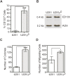
Figure 3. Bioenergetic characterization of mtDNA depleted U251 glioma cells.
A) Genomic DNA was isolated from control and mtDNA-depleted U251 cells, and mtDNA-encoded genes including cytochrome c oxidase subunit I (COX-I) and cytochrome c oxidase subunit II (COX-II) were amplified by PCR. COX IV is used as a nuclear DNA-encoded control. The PCR products were electrophoresed on 1.5% agarose gel and then visualized by EtBr staining. A representative result of three independent experiments is shown. B) Average oxygen consumption rates in U251 and U251ρ0. Results are given as means ± SD of 5 separate experiments. C) Rhodamine123 fluorescence profiles of U251 and U251ρ0; NAO fluorescent profiles of U251 and U251ρ0. D) Glucose transport activity for the parental and ρ0 cells. Results are given as means ± SD of 3 separate experiments.
Figure 4
Figure 4. Anchorage-independent, invasion and CD133 expression in ρ0 glioma cells.
A) Flow cytometry determination of the percentage of CD133 expressing parental and ρ0 cells. Results are given as means ± SD of 5 separate experiments. B) RT-PCR detection of CD133 mRNA from U251 and U251ρ0 glioma cells. Electrophoresis resolved amplicons for CD133 (0.4 kbp) and actin (0.2 kbp) of anticipated sizes. C) Colony formation in soft agar. Results are given as means ± SD of 3 separate experiments. D) Analysis of invasion into Matrigel 24 h after 5% FCS as chemo-attractant has been added to the lower chamber. *, **, *** represent p<0.05, p<0.005 and p<0.001.
Figure 5
Figure 5. ρ0 glioma cells form neurosphere-like tumor spheroids expressing neural stem cell markers, nestin and CD133.
A) Phase contrast microphotographs (magnification X20) of ρ0 glioma forming (a) neurosphere-like structures in serumless Neurobasal medium supplement with EGF and FGF after 10 days in culture or (b) adherent culture in DMEM/F12 medium with 7% FBS. B) Spheroids of ρ0 cells were immunostained with (b) CD133 or (c) Nestin antibodies or nuclear counterstained with Hoechst 33258 (a). C) Differentiation potential of ρ0 tumor spheroids. Multipotency was assayed by immunofluorescence for neuronal (βIII-tubulin) (a, c) and glial (GFAP) (b a, c) markers.
Figure 6
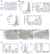
Figure 6. Characterization of U251 transmitochondrial cybrids.
A) Genomic DNA was isolated from control, mtDNA-depleted and transmitochondrial cybrid cells, and mtDNA-encoded genes including COX-I and COX-II were amplified by PCR. COX IV serves as a nuclear DNA-encoded control. The PCR products were electrophoresed on 1.5% agarose gel and then visualized by EtBr staining. A representative result of three independent experiments is shown. B) Representative histograms of R123 fluorescence for U251, U251ρ0 and cybrids cells (left); average R123 fluorescence profiles (right). C) Representative histograms of NAO fluorescence for U251, U251ρ0 and cybrids cells (left); average NAO fluorescence profiles (right). D) A Representative electron transmission microscopy photographs comparing U251, U251ρ0 and cybrids cells (Magnification X6000). Arrowhead showing that U251ρ0 cells have smaller mitochondria with large intramembraneous spaces. E) Glucose transport activity for the parental and ρ0 cells. Results are given as means ± SD of 3 separate experiments. F) Representative histograms of flow cytometry CD133 fluorescence for U251, U251ρ0 and cybrids cells (left). Percentage of CD133 expressing cells (right). Results are given as means ± SD of 5 separate experiments.
Figure 7
Figure 7. Tumor progression model:
(a) Initiation of tumor formation. (b) Tumor growth restricted at the oxygen and glucose diffusion barrier. (c) Glioma ells at the edge of the tumor will be re-oxygenated by neovascularization, and grow and divide until they reach a new oxygen and nutrient diffusion barrier. (d): repetitive cycles of growth and angiogenesis occur during the progression of the tumor.

References

    1. Griguer CE, Oliva CR, Gillespie GY. Glucose metabolism heterogeneity in human and mouse malignant glioma cell lines. J Neurooncol. 2005;74:123–133. - PubMed
    1. Griguer CE, Oliva CR, Gillespie GY, Gobin E, Marcorelles P, et al. Pharmacologic manipulations of mitochondrial membrane potential (DeltaPsim) selectively in glioma cells. J Neurooncol. 2007;81:9–20. - PubMed
    1. Griguer CE, Oliva CR, Kelley EE, Giles GI, Lancaster JR, Jr, et al. Xanthine oxidase-dependent regulation of hypoxia-inducible factor in cancer cells. Cancer Res. 2006;66:2257–2263. - PubMed
    1. Warburg O. On respiratory impairment in cancer cells. Science. 1956;124:269–270. - PubMed
    1. Modica-Napolitano JS, Kulawiec M, Singh KK. Mitochondria and human cancer. Curr Mol Med. 2007;7:121–131. - PubMed

Publication types

MeSH terms