

The Role of Aromatic Residues in Stabilizing the Secondary and Tertiary Structure of Avian Pancreatic Polypeptide
- PMID: 18985166
- PMCID: PMC2577375
- DOI: 10.1002/qua.21521
The Role of Aromatic Residues in Stabilizing the Secondary and Tertiary Structure of Avian Pancreatic Polypeptide
Abstract
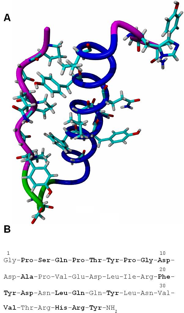
Avian Pancreatic Polypeptide is a 36 residue protein that exhibits a tertiary fold. Results of previous experimental and computational studies indicate that the structure of aPP is stabilized more by non-bonded interactions than by the hydrophobic effect. Aromatic residues are known to participate in a variety of long range non-bonded interactions, with both backbone atoms and the atoms of other side-chains, which could be responsible, in part, for the stability of both the local secondary structure and the tertiary fold. The effect of these aromatic interactions on the stability of aPP was calculated using BHandHLYP/cc-pVTZ. Aromatic residues were shown to participate in multiple hydrogen bonded and weakly polar interactions in the secondary structure. The energies of the weakly polar interactions are comparable with those of hydrogen bonds. Aromatic residues were also shown to participate in multiple weakly polar interactions across the tertiary fold, again with energies similar to those of hydrogen bonds.
Figures


References
-
- Covell DG, Jernigan RL. Biochemistry. 1990;29:3287–3294. - PubMed
Grants and funding
LinkOut - more resources
Full Text Sources