. 2006 May;2(3):649-655.
doi: 10.1021/ct050298p.

Probing Membrane Insertion Activity of Antimicrobial Polymers via Coarse-grain Molecular Dynamics

Affiliations

Probing Membrane Insertion Activity of Antimicrobial Polymers via Coarse-grain Molecular Dynamics

Carlos F Lopez et al. J Chem Theory Comput. 2006 May.

Abstract

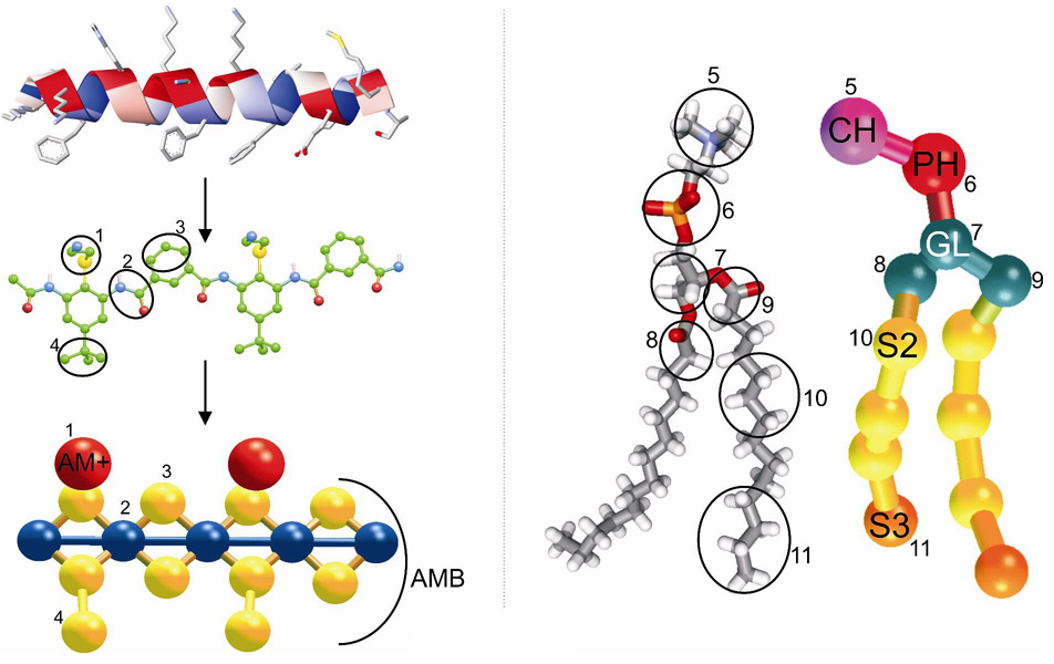
Knowledge of the mechanism of action of antimicrobial agents is crucial for the development of new compounds to combat microbial pathogens. To this end, computational studies on the interaction of known membrane-active antimicrobial polymers with phospholipid bilayers reveal spontaneous membrane insertion and cooperative action at low and high concentrations, respectively. In late-stage attack, antimicrobials cross the membrane core and occasionally align to provide a stepping-stone pathway for water permeation; this suggests a possible new mode of action that does not depend on pore formation for transport to and across the inner leaflet. The computations rationalize the observed activity of a new class of antimicrobial compounds.


Figures

Figure 1
Figure 1
Inspiration for the design of synthetic antimicrobial targets and the coarse grain model used in the present simulations. LEFT: Ribbon representation of magainin, a potent natural antimicrobial. The α-helix ribbon is colored by hydrophobicity (red=hydrophobic, blue=hydrophilic), and selected individual amino acids are shown as sticks. Middle: The aryl amide dimeric polymer, a purely synthetic molecule inspired by natural antimicrobials. Color code: carbon, green; sulphur, yellow; oxygen, red; nitrogen, blue; hydrogen, white. Bottom: A coarse grain representation of the aryl amide dimeric polymer. Color code: cationic, red; peptidic, blue; hydrophobic, yellow. RIGHT: The corresponding units between an all atom DMPC molecule (left) and its CG representation (right). Color code: nitrogen, blue; carbon, grey; oxygen, red; phosphate, orange; hydrogen, white. CG color code: choline (CH), purple; phosphate (PH), red; glycerol (GL), blue-gray; alkane (S2), yellow; terminal alkane (S3), orange. Circles in the atomistic representations correspond to the numbered units in the coarse grain representations.
Figure 2
Figure 2
Depiction of the observed two-step - accommodation (a) and penetration (b) mechanism for membrane insertion of the antimicrobial (AM) aryl amide dimer molecules into the outer leaflet of a DMPC lipid bilayer (DMPC: choline, red; phosphate, purple; glycerol group, blue sticks; hydrophobic tails, green sticks). The initial interfacial insertion stage involves adsorption and “snorkeling” of the AM at the bilayer surface (a). In this example of the penetration stage, one AM rotates to become perpendicular to the bilayer plane and drags the accompanying AM into the membrane core (b). Subsequent population of both leaflets occurs at much longer times (c).
Figure 3
Figure 3
Insertion distance to bilayer center (top graphs) and orientation angle (bottom graphs) for the 1AM simulation (left) and the 2AM simulation (right). Dashed lines represent the average positions of the CH group (top line) and the GL group (bottom line) within the attacked leaflet.
Figure 4
Figure 4
Atom weighted densities for the lipid bilayer (solid lines) and the AM molecules (dashed lines) for simulations with none (black), two (red), eight (green), and eighteen (blue) antimicrobial molecules. The scale on the left corresponds to the lipid molecules while the scale on the right corresponds to the AM polymers.
Figure 5
Figure 5
Atom weighted densities for the lipid bilayer (solid lines) and the AM molecules (dashed lines) for simulations with none (black), two (red), eight (green), and eighteen (blue) antimicrobial molecules. The scale on the left corresponds to the lipid molecules while the scale on the right corresponds to the AM polymers.
Figure 6
Figure 6
Representative water trajectories (blue spheres are CG waters) showing passage from the bulk water phase, W1 (purple), through the head group region HG1 (orange), and hydrophobic membrane core, C (yellow) to the HG2 and W2 regions via a pathway employing the aryl amide molecules as stepping stones.

Similar articles

Cited by

References

    1. Brower V. EMBO Rep. 2003;4:649–651. - PMC - PubMed
    1. Cohen ML. Nature. 2000;406:762–767. - PubMed
    1. Anderson RM. Nature Med. 1999;5:147–149. - PubMed
    1. Niller E. Nature Biotechnol. 2002;20:21–25. - PubMed
    1. Fernandez-Lopez S, Kim HS, Choi EC, Delgado M, Granja JR, Khasanov A, Kraehenbuehl K, Long G, Weinberger DA, Wilcoxen KM, Ghadiri MR. Nature. 2001;412:452–455. - PubMed

LinkOut - more resources