Case Reports
. 2009 Jun;118(3):239-46.
doi: 10.1007/s10633-008-9154-5. Epub 2008 Nov 5.

Detailed analysis of retinal function and morphology in a patient with autosomal recessive bestrophinopathy (ARB)

Affiliations
Case Reports

Detailed analysis of retinal function and morphology in a patient with autosomal recessive bestrophinopathy (ARB)

Christina Gerth et al. Doc Ophthalmol. 2009 Jun.

Abstract

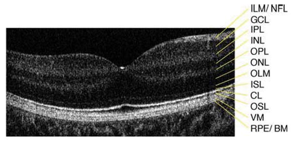
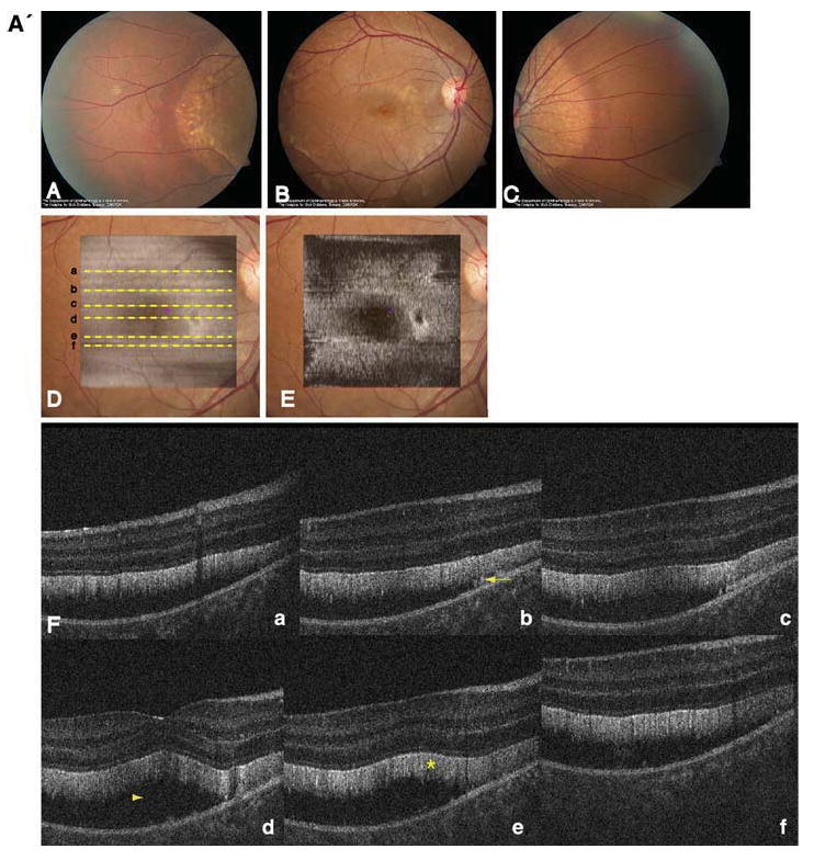
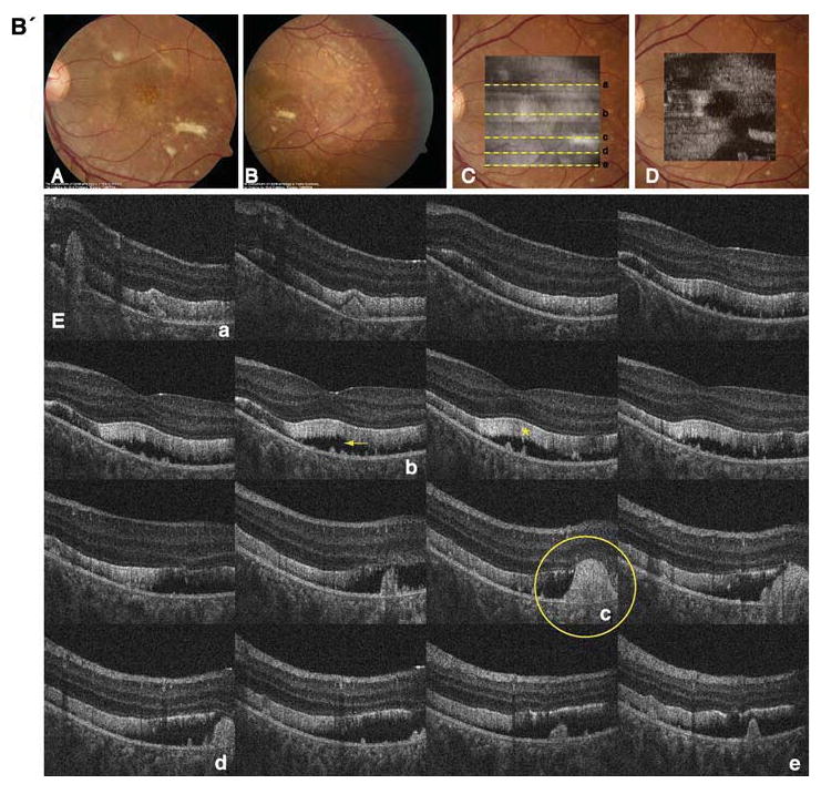
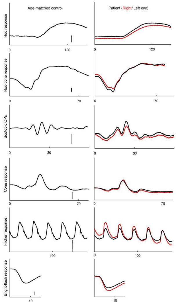
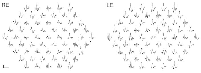
The objective of the paper is to study the retinal microstructure and function in a patient with autosomal recessive bestrophinopathy (ARB). Retinal function and morphology assessment in a patient diagnosed with a biallelic mutation in the BEST1 gene (heterozygote mutations: Leu88del17 and A195V) included: full-field electroretinogram (ffERG) and multifocal electroretinogram (mfERG), electro-oculogram (EOG) testing, and imaging with a high-resolution Fourier-domain optical coherence tomography (Fd-OCT) system (UC Davis Medical Center; axial resolution: 4.5 microm, acquisition speed: 9 frames/s, 1,000 A-scans/frame) combined with a flexible scanning head (Bioptigen Inc.). The 11-year old asymptomatic boy showed a well-demarcated retinopathy with deposits. Functional assessment revealed normal visual acuity, reduced central mfERG responses, delayed rod and rod-cone b-wave ffERG responses, and reduced light rise in the EOG. Fd-OCT demonstrated RPE deposits, photoreceptor detachment, elongated and thickened photoreceptor outer segments, but preserved inner retinal layers. In conclusion, ARB associated retinal dystrophy shows functional and morphological changes that overlap with classic Best disease. For the first time, high-resolution imaging provided in vivo evidence of RPE and photoreceptor involvement in ARB.


Figures

Fig. 1
Fig. 1
Six millimeter horizontal Fd-OCT scan through the right macula of a 9.3 year-old control subject. CL, connecting cilia; GCL, ganglion cell layer; ILM/NFL, internal limiting membrane/nerve fiber layer; INL, inner nuclear layer; IPL, inner plexiform layer; ISL, inner segment layer; OLM, outer limiting membrane; ONL, outer nuclear layer; OPL, outer plexiform layer; OSL, outer segment layer; RPE/BM, retinal pigment epithelium/Bruch's membrane; VM, Verhoeff's membrane
Fig. 2
Fig. 2
A' Composite of color fundus photographs and serial horizontal Fd-OCT scans (6 mm) of the right eye. Fundus photograph shows a well-demarcated area with round yellowish-white deposits at the posterior pole (A) extending to the temporal (B) and nasal (C) periphery. The locations of OCT B-scans are denoted by lines (af) shown on the OCT fundus image (intensity projection of the OCT volume), which is superposed and registered to the color fundus photo. (D) Virtual C-scan at the level of the photoreceptor inner/outer segment junction segmented from the reconstructed OCT volume is shown. (E) B-scans (F) illustrate deposits (denoted by an arrow in one scan) within the RPE with RPE detachment from the photoreceptors (arrowhead in one scan). Photoreceptor outer segments are thickened and elongated (indicated by a star in one scan). B' Composite of color fundus photograph and serial horizontal Fd-OCT scans (6 mm) of the left eye. Fundus photograph shows a well-demarcated area with round yellowish-white deposits at the posterior pole (A) extending to the temporal (B) and nasal (not shown) periphery. Subretinal fibrosis is visible inferior-temporal and superior-nasal of the fovea. The locations of OCT B-scans are denoted by lines (examples ae) shown on the OCT fundus image, which is superposed and registered to the color fundus photo. (C) Virtual C-scan at the level of the photoreceptor inner/outer segment junction segmented from the reconstructed OCT volume is shown. (D) B-scans (E) demonstrate RPE detachment from the photoreceptors. Photoreceptor outer segments are thickened and elongated (indicated by a star in one scan). Small bridges between the photoreceptor outer segments and RPE are visible as denoted by an arrow in the scan through the fovea (b). Circle illustrates extensive RPE deposits extending to the outer plexiform layer
Fig. 2
Fig. 2
A' Composite of color fundus photographs and serial horizontal Fd-OCT scans (6 mm) of the right eye. Fundus photograph shows a well-demarcated area with round yellowish-white deposits at the posterior pole (A) extending to the temporal (B) and nasal (C) periphery. The locations of OCT B-scans are denoted by lines (af) shown on the OCT fundus image (intensity projection of the OCT volume), which is superposed and registered to the color fundus photo. (D) Virtual C-scan at the level of the photoreceptor inner/outer segment junction segmented from the reconstructed OCT volume is shown. (E) B-scans (F) illustrate deposits (denoted by an arrow in one scan) within the RPE with RPE detachment from the photoreceptors (arrowhead in one scan). Photoreceptor outer segments are thickened and elongated (indicated by a star in one scan). B' Composite of color fundus photograph and serial horizontal Fd-OCT scans (6 mm) of the left eye. Fundus photograph shows a well-demarcated area with round yellowish-white deposits at the posterior pole (A) extending to the temporal (B) and nasal (not shown) periphery. Subretinal fibrosis is visible inferior-temporal and superior-nasal of the fovea. The locations of OCT B-scans are denoted by lines (examples ae) shown on the OCT fundus image, which is superposed and registered to the color fundus photo. (C) Virtual C-scan at the level of the photoreceptor inner/outer segment junction segmented from the reconstructed OCT volume is shown. (D) B-scans (E) demonstrate RPE detachment from the photoreceptors. Photoreceptor outer segments are thickened and elongated (indicated by a star in one scan). Small bridges between the photoreceptor outer segments and RPE are visible as denoted by an arrow in the scan through the fovea (b). Circle illustrates extensive RPE deposits extending to the outer plexiform layer
Fig. 3
Fig. 3
Full-field electroretinogram of the patient's right (red traces) and left eye (black traces) and an age-matched control (amplitude scale bar: 100 μV except OPs: 50 μV). The patient's rod and rod-cone b-wave responses are delayed in both eyes. (Table 1)
Fig. 4
Fig. 4
Multifocal electroretinogram traces from the right and left eye are shown. Responses within the central 10° are reduced (scale bar 200 nV, 80 ms)

References

    1. Burgess R, Millar ID, Leroy BP, et al. Biallelic mutation of BEST1 causes a distinct retinopathy in humans. Am J Hum Genet. 2008;82:19–31. - PMC - PubMed
    1. Petrukhin K, Koisti MJ, Bakall B, et al. Identification of the gene responsible for Best macular dystrophy. Nat Genet. 1998;19:241–247. - PubMed
    1. Stohr H, Marquardt A, Rivera A, et al. A gene map of the Best's vitelliform macular dystrophy region in chromosome 11q12–q13.1. Genome Res. 1998;8:48–56. - PMC - PubMed
    1. Marquardt A, Stohr H, Passmore LA, et al. Mutations in a novel gene, VMD2, encoding a protein of unknown properties cause juvenile-onset vitelliform macular dystrophy (Best's disease) Hum Mol Genet. 1998;7:1517–1525. - PubMed
    1. Marmor MF, Holder GE, Seeliger MW, Yamamoto S. Standard for clinical electroretinography (2004 update) Doc Ophthalmol. 2004;108:107–114. - PubMed

Publication types

MeSH terms