Solitary fibrous tumor of the liver expressing CD34 and vimentin: a case report
- PMID: 18985821
- PMCID: PMC2761592
- DOI: 10.3748/wjg.14.6261
Solitary fibrous tumor of the liver expressing CD34 and vimentin: a case report
Abstract
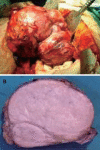
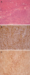
A case of a successfully treated solitary fibrous tumor (SFT) of the liver is reported. An 82-year-old female presented with left upper abdominal discomfort, a firm mass on palpation, and imaging studies revealed a large tumor, 15 cm in diameter, arising from the left lobe of the liver. A formal left hepatectomy was performed. Microscopic evaluation showed spindle and fibroblast-like cells within the collagenous stroma. Immunohistochemistry disclosed diffuse CD34 and positive vimentin, supporting the diagnosis of a benign SFT. The patient remained well 21 months after surgery. SFT of the liver is a very rare neoplasm of mesenchymal origin. In most cases it is a benign lesion, although some may have malignant histological features and recur locally or metastasize. With less than 30 reported cases in the literature, little can be said regarding its natural history or the benefits of adjuvant radiochemotherapy. Complete surgical resection remains the cornerstone of its treatment.
Figures


References
-
- Neeff H, Obermaier R, Technau-Ihling K, Werner M, Kurtz C, Imdahl A, Hopt UT. Solitary fibrous tumour of the liver: case report and review of the literature. Langenbecks Arch Surg. 2004;389:293–298. - PubMed
-
- Changku J, Shaohua S, Zhicheng Z, Shusen Z. Solitary fibrous tumor of the liver: retrospective study of reported cases. Cancer Invest. 2006;24:132–135. - PubMed
-
- Vennarecci G, Ettorre GM, Giovannelli L, Del Nonno F, Perracchio L, Visca P, Corazza V, Vidiri A, Visco G, Santoro E. Solitary fibrous tumor of the liver. J Hepatobiliary Pancreat Surg. 2005;12:341–344. - PubMed
-
- Ji Y, Fan J, Xu Y, Zhou J, Zeng HY, Tan YS. Solitary fibrous tumor of the liver. Hepatobiliary Pancreat Dis Int. 2006;5:151–153. - PubMed
-
- Guglielmi A, Frameglia M, Iuzzolino P, Martignoni G, De Manzoni G, Laterza E, Veraldi GF, Girlanda R. Solitary fibrous tumor of the liver with CD 34 positivity and hypoglycemia. J Hepatobiliary Pancreat Surg. 1998;5:212–216. - PubMed
Publication types
MeSH terms
Substances
LinkOut - more resources
Full Text Sources
Medical

