. 2008 Nov 5:3:30.
doi: 10.1186/1749-8104-3-30.

Genetic lineage tracing defines distinct neurogenic and gliogenic stages of ventral telencephalic radial glial development

Affiliations

Genetic lineage tracing defines distinct neurogenic and gliogenic stages of ventral telencephalic radial glial development

Todd E Anthony et al. Neural Dev. .

Abstract

Background: Radial glia comprise a molecularly defined neural progenitor population but their role in neurogenesis has remained contested due to the lack of a single universally accepted genetic tool for tracing their progeny and the inability to distinguish functionally distinct developmental stages.

Results: By direct comparisons of Cre/loxP lineage tracing results obtained using three different radial glial promoters (Blbp, Glast, and hGFAP), we show that most neurons in the brain are derived from radial glia. Further, we show that hGFAP promoter induction occurs in ventral telencephalic radial glia only after they have largely completed neurogenesis.

Conclusion: These data establish the major neurogenic role of radial glia in the developing central nervous system and genetically distinguish an early neurogenic Blbp+Glast+hGFAP- stage from a later gliogenic Blbp+Glast+hGFAP+ stage in the ventral telencephalon.


Figures

Figure 1
Figure 1
Glast bacterial artificial chromosome (BAC) transgenic mice recapitulate the spatiotemporal pattern of endogenous GLAST (glial high affinity glutamate transporter) expression. (e-v") In situ hybridization (ISH) (a-d) and immunofluorescent staining (e-v) of Glast::Cre (a-i) and Glast::eGFP (j-v) BAC transgenic mice. Cre mRNA in the ganglionic eminences (a), thalamus (b), spinal cord (c), and cerebellum/hindbrain (d) was completely restricted to the ventricular zone, with no neuronal expression detected at any age examined. The Glast::Cre transgene also recapitulated endogenous GLAST expression gradients (compare (c) and (r)). Postnatally, Cre immunoreactivity (green) (e-i) was restricted to astroglia. Shown are cortical GLAST+ astrocytes (f), striatal BLBP+ astrocytes (g), and cerebellar Bergmann and astroglia (h,i); no colocalization with CALB1+ Purkinje cells was observed (i). Double immunofluorescent staining of Glast::eGFP embryos for GLAST (red) and enhanced green fluorescent protein (eGFP; green) showed that the BAC transgene drives expression in essentially all GLAST+ radial glia (j-q); the notable exception to this was the spinal cord floorplate where no transgene expression was detectable (r-t). Postnatal expression was similarly restricted to astroglia (u.v). Scale bars: 200 μm (c,d,e,h,r,s); 100 μm (b); 75 μm (a,j-l); 50 μm (f,n-p,t); 40 μm (m); 35 μm (u); 25 μm (g,i,q,v). E, embryonic day.
Figure 2
Figure 2
Recombination in Glast::Cre;R26R embryos parallels endogenous GLAST (glial high affinity glutamate transporter) expression and the pattern observed in Blbp::Cre;R26R embryos. (a-q) Wild-type (a-f), Glast::Cre;R26RlacZ (g-m) and Glast::Cre;Z/EG (n-q) embryos immunostained for GLAST (a,b,e,f,k,l), BLBP (c-f), phosphorylated histone H3 (PH3) (n,o,q) or GFP (p,q) and X-gal staining (g-j,k',l',m'). Sections in (a-j) are sagittal; (k-q) are coronal. At E9.5, endogenous GLAST (a) and BLBP (c) expression was detected in hindbrain and spinal cord, but not in telencephalon until E10.5 (b,d). Double labeling for GLAST (red) and BLBP (green) in E10.5 CX and GE confirmed that both are upregulated in the same cells (e,f). The onset of Cre recombination matched the spatiotemporal pattern of endogenous GLAST expression, with minimal β-Gal activity in E9.5 telencephalon (g,h) but extensive staining by E10.5 (i,j); this pattern matches that previously reported for Blbp::Cre;R26R embryos [8]. Concordance between β Gal activity and GLAST+ RG was consistently observed. At E12.5, GLAST is weak in septum (k'), and recombination in this region was similarly minimal (k). Conversely, GLAST and recombination are intense in the cortical hem (arrow in (l); higher magnification image of GLAST hem staining in (l')). Further, both Cre mRNA 1c) and recombination (m) in the spinal cord matched the dorsoventral gradient of endogenous GLAST in this region (Figure 1r). PH3 and GFP labeling in the E14.5 GE of Glast::Cre;Z/EG embryos showed that mitotic SVZ progenitors were recombined (n-q). (as observed in Blbp::Cre;R26R embryos [8], establishing that GE SVZ progenitors are RG-derive. Scale bars: 400 μm (a,c,g,i); 200 μm (k); 175 μm (b,d,j); 150 μm (h); 100 μm (m); 40 μm (f,n); 25 μm (l); 20 μm (e,o-q).
Figure 3
Figure 3
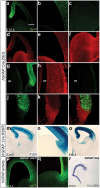
Glast+ radial glia generate the majority of neurons in the brain. (a-o) X-gal histochemical staining for β-gal activity (a,b,f,g,k,l) and immunofluorescent staining for NeuN (red) (c,e,h,j), β-galactosidase (green; d,e,i,j,n,o) and Calbindin (red) (m,o) in hGFAP::Cre;R26R (a,f,k) or Glast::Cre;R26R (b-e,g-j,l-o) adult mice. Regions immunofluorescently stained are striatum (c-e), thalamus (h-j) and cerebellum (m-o). In hGFAP::Cre;R26R mice, very few recombined neurons are present in the ventral telencephalon, diencephalon or midbrain, and cerebellar Purkinje neurons are unrecombined (a,f,k). PCL, Purkinje cell layer. In contrast, neuronal recombination was extensive throughout the brains of Glast::Cre;R26R mice (b,g,l) and double labeling confirmed that most neurons were recombined (c-e,h-j,m-o). The similarity of the Glast::Cre;R26R and Blbp::Cre;R26R lineage tracing results establishes that radial glia are the primary neuronal progenitors throughout the developing brain. Scale bars: 500 μm (a,b,f,g); 50 μm (k,l); 35 μm (m-o); 25 μm (c-e,h-j).
Figure 4
Figure 4
The hGFAP promoter fails to drive recombination in fully morphologically differentiated radial glia (RG) of the ventral telencephalon and thalamus. Immunostaining for eGFP (green) (a-c,g,i,j,l,p,q) and GLAST (red) (d-f,h,k,l), X-gal staining for β-gal (m-o) and Cre ISH (r) on hGFAP::Cre;Z/EG (a-l), hGFAP::Cre;R26R (m-o,r) or hGFAP::eGFP (p,q) embryos, at E12.5 (a-l), E14.5 (m,n,p,q), E15.5 (o), and E16.5 (r). Panels (b,c,e,f) show cortex and GE in (a,d) at higher magnification; panels (h,i) show thalamus in (g) at higher magnification, arrows in (g-i) mark identical spots in all three panels; panels (n,q) show higher magnification images of cortex and GE in (m,p). In contrast to the Blbp and Glast promoters, the hGFAP promoter is not active in forebrain prior to E12.5 (not shown). By E12.5, recombination is detected in cortical RG but absent from GE (a-c), thalamus (g,i) and cortical hem (j,l). Importantly, both BLBP (not shown) and GLAST (d-f,h,k,l) are highly expressed in these regions. Moreover, these unrecombined cells have molecular and morphological properties of fully differentiated RG cells (e.g. long radial processes coursing from VZ to pia). Even as late as E14.5, few GE RG are recombined (m,n), and significant recombination is not observed until E15.5 (o) when most RG in the region have completed neurogenesis. hGFAP promoter activity itself is not only delayed with respect to RG differentiation, but also weak ventrally compared to its dorsal activity (p,q,r). Scale bars: 300 μm (m,o,r); 200 μm (a,d,p); 100 μm (c,f,g,n,q); 60 μm (b,e,h-l).

References

    1. Rakic P. Elusive radial glial cells: historical and evolutionary perspective. Glia. 2003;43:19–32. doi: 10.1002/glia.10244. - DOI - PubMed
    1. Kriegstein AR. Constructing circuits: neurogenesis and migration in the developing neocortex. Epilepsia. 2005;46:15–21. doi: 10.1111/j.1528-1167.2005.00304.x. - DOI - PubMed
    1. Noctor SC, Flint AC, Weissman TA, Dammerman RS, Kriegstein AR. Neurons derived from radial glial cells establish radial units in neocortex. Nature. 2001;409:714–720. doi: 10.1038/35055553. - DOI - PubMed
    1. Malatesta P, Hartfuss E, Gotz M. Isolation of radial glial cells by fluorescent-activated cell sorting reveals a neuronal lineage. Development. 2000;127:5253–5263. - PubMed
    1. Malatesta P, Hack MA, Hartfuss E, Kettenmann H, Klinkert W, Kirchhoff F, Gotz M. Neuronal or glial progeny: regional differences in radial glia fate. Neuron. 2003;37:751–764. doi: 10.1016/S0896-6273(03)00116-8. - DOI - PubMed

Publication types

MeSH terms

Substances

LinkOut - more resources