

doi: 10.1093/hmg/ddn358.
Epub 2008 Nov 4.
Myogenic Akt signaling upregulates the utrophin-glycoprotein complex and promotes sarcolemma stability in muscular dystrophy
Affiliations
- PMID: 18986978
- PMCID: PMC2638781
- DOI: 10.1093/hmg/ddn358
Item in Clipboard
Myogenic Akt signaling upregulates the utrophin-glycoprotein complex and promotes sarcolemma stability in muscular dystrophy
Hum Mol Genet.
.
Abstract
Duchenne muscular dystrophy is caused by dystrophin mutations that lead to structural instability of the sarcolemma membrane, myofiber degeneration/regeneration and progressive muscle wasting. Here we show that myogenic Akt signaling in mouse models of dystrophy promotes increased expression of utrophin, which replaces the function of dystrophin thereby preventing sarcolemma damage and muscle wasting. In contrast to previous suggestions that increased Akt in dystrophy was a secondary consequence of pathology, our findings demonstrate a pivotal role for this signaling pathway such that modulation of Akt can significantly affect disease outcome by amplification of existing, physiological compensatory mechanisms.
Figures
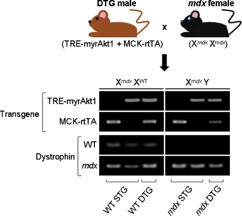
Generation of Akt-transgenic mdx mice. Breeding scheme and genotyping of offspring by PCR amplification. Double-transgenic (TRE-myrAkt1 + MCK-rtTA) male mice on a wild-type (WT) dystrophin background were crossed with mdx females. Each transgenic offspring inherited either one or both of the Akt transgenes (TRE-myrAkt1, MCK-rtTA). All female offspring, which were heterozygous for dystrophin (Xmdx XWT) and expressing non-dystrophic phenotypes, are denoted as being either wild-type single-transgenic (WT STG) or wild-type double-transgenic (WT DTG). All male offspring possessed mdx genotypes (Xmdx Y) and are denoted as being either mdx STG or mdx DTG. Transgenic offspring were genotyped through two pairs of PCR amplification reactions. In the first pair of reactions, offspring were genotyped for the two transgenes responsible for the activation of constitutively active Akt1. In the first PCR reaction, the transgene for the constitutively active form of Akt1 under the control of a tetracycline responsive promoter (TRE-myrAkt1) is amplified, yielding a 380 bp product. In the second PCR reaction, the transgene expressing a reverse tetracycline transactivator under the control of a modified muscle creatine kinase promoter (MCK-rtTA) was amplified, yielding a 567 bp product. In the second pair of reactions, offspring were genotyped for the mdx locus. The forward primers used in this pair of reactions are identical. In the first reaction of the pair, the WT allele was amplified using a WT allele-specific reverse primer. In the second reaction, the mdx allele was amplified using an mdx allele-specific reverse primer. Each reaction in this pair yielded a 275 bp product.
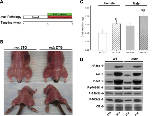
Akt1 activation increases muscle mass in mdx mice. (A) Timeline of doxycycline (DOX) treatment of Akt STG and DTG mice. At three weeks of age, the mice were treated with DOX, inducing Akt expression in Akt DTG mice for a total of three weeks. DOX treatment period corresponds to period of maximal myofiber degeneration and regeneration in mdx mice. Tissues were analyzed at six weeks of age. (B) Representative images of mdx STG and mdx DTG mice reveal dramatic differences in muscle mass. DTG mice display increased muscle mass (asterisk) and hypervascularization (arrow) after three weeks of transgene activation. (C) Activation of Akt significantly increases muscle mass in DTG mice. Induction of transgene in DTG mice significantly increases total quadriceps weight when compared to their WT (Fisher's, *P < 0.00001) and mdx STG (Fisher's, **P < 0.005) counterparts. Quadriceps weights are represented as an average of the left and right quadriceps of each animal. Bars represent mean quadriceps weights (±SEM; n = 21 mdx STG, n = 4 mdx DTG. n = 18 WT STG, n = 11 WT DTG). (D) Immunoblotting for Akt pathway proteins. Akt pathway proteins were detected from total skeletal muscle lysates of six-week-old WT STG, WT DTG, mdx STG, and mdx DTG mice. Identical membranes were probed with antibodies against Akt, P-Akt, P-70S6K, P-GSK3β, P-MDM2, and the HA-tag engineered onto the Akt1 transgene, as indicated. Coomassie blue staining of total protein is shown on the bottom panel (CB Stain) as a loading control. Activation of Akt for three weeks is sufficient for activation of P-70S6K in the Akt signaling axis. Furthermore, P-MDM2 levels increase upon Akt activation whereas P-GSK3β levels remain constant.
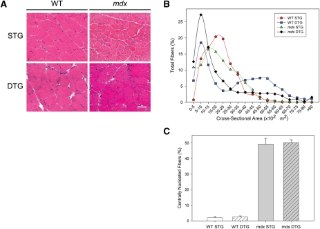
Akt1 activation increases fiber hypertrophy. (A) Transverse quadriceps muscle sections from six-week-old WT STG, WT DTG, mdx STG, and mdx DTG mice were stained with hematoxylin and eosin (H&E) to visualize muscle histology. Constitutive Akt activation in mdx DTG mice did not reduce the prevalence of centrally nucleated fibers and necrosis when compared to non-phenotypic littermates. Note the muscle fiber hypertrophy in both groups of DTG mice. Bar, 50 μm. (B) Distribution of cross sectional fiber areas of quadriceps muscle. DTG mice exhibit greater populations of both smaller and hypertrophic fibers compared to their WT and mdx STG controls. (C) Central nucleation (% of total fibers) in quadriceps muscle sections was quantified in each group of mice. Mdx mice exhibit significantly higher levels of central nucleation when compared to WT mice (*ANOVA, P < 0.005). However, DTG mice do not exhibit significant differences in the amount of central nucleation when compared to their STG control littermates. Central nucleation is represented as an average of the % of central nucleation of the left and right quadriceps of each animal. Bars represent mean central nucleation. (n = 3 WT STG, n = 2 WT DTG, n = 2 mdx STG, n = 3 mdx DTG.).
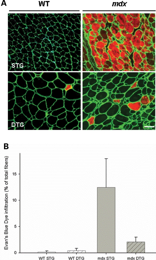
Akt1 activation ameliorates sarcolemmal stability in mdx DTG mice. (A) Detection of blood serum protein infiltration with Evans Blue dye (EBD) tracer assay. Transverse sections of quadriceps muscle fibers with damaged sarcolemma are shown with red fluorescence. Laminin in the basement membrane is shown with green fluorescence to delineate each myofiber. EBD infiltration was absent in WT STG, detected in low levels in WT DTG and mdx DTG mice, and elevated in mdx STG mice. Bar, 50 μm. (B) Quantification of EBD-positive fibers in whole transverse quadriceps sections. Bars represent percentage of EBD infiltrated fibers in a whole quadriceps section (±SEM; Student's t-test with Bonferroni adjustment (α = 0.025), N.S. WT STG versus WT DTG, *P < 0.02, mdx DTG versus mdx STG; n = 3 mice).
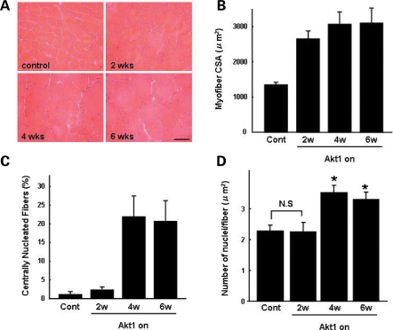
Akt activates muscle regeneration (A) Adult WT DTG mice were treated with DOX for two, four and six weeks. Transverse gastrocnemius muscle sections from treated WT DTG mice were stained with hematoxylin and eosin (H&E) to visualize muscle histology. Muscle sections taken from mice treated with vehicle control is shown (control). Note that muscle fiber hypertrophy is evident after two weeks of DOX administration. Bar, 50 μm. (B) Cross-sectional area (CSA) of transverse gastrocnemius WT muscle was quantified after two, four and six weeks of DOX treatment. Mean values (μm2) are provided to illustrate overall myofiber hypertrophy that is evident after two weeks of Akt1 transgene activation. Values from control, vehicle-treated muscle are shown. (C) Central nucleation (% of total fibers) in gastrocnemius muscle sections was quantified in each group of mice. Regeneration, denoted by the presence of myofibers with centrally placed nuclei, is evident in WT muscle after four weeks of Akt activation. This regeneration is occurring in the absence of muscle pathology or disease. Central nucleation is represented as an average of the % of central nucleation. (D) Quantification of the number of nuclei per myofiber is provided. No statistical (N.S.) difference in nuclei per myofiber was observed after two weeks of DOX treatment. After four and six weeks of Akt activation, the number of nuclei/myofiber increased by ∼50% relative to controls. In panels B-D, bars represent standard error of the mean (Fisher's *P < 0.05; n = 7).
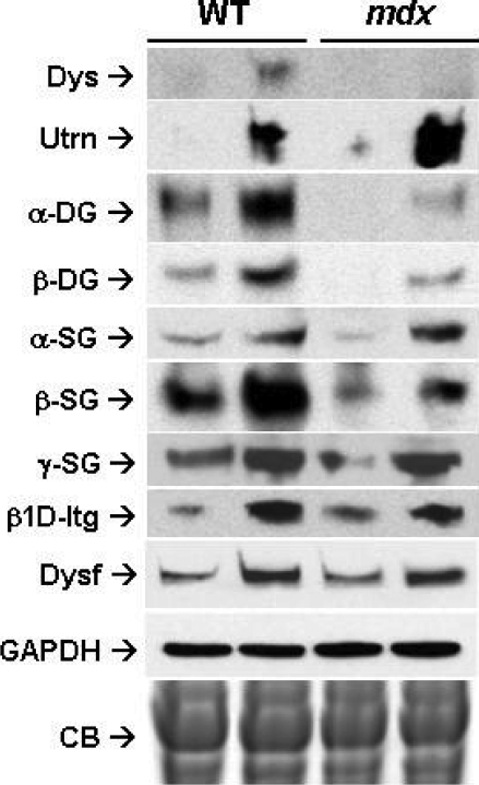
Akt increases expression of compensatory proteins in mdx mice. Immunoblotting for several glycoprotein complexes (DGC, UGC, integrin) on skeletal muscle lysates from six-week-old WT STG, WT DTG, mdx STG, and mdx DTG mice. Identical membranes were probed with antibodies against dystrophin (Dys), utrophin (Utrn), alpha- and beta-dystroglycan (α-DG, β-DG), alpha-, beta- and gamma-sarcoglycan (α-SG, β-SG, γ-SG), β1D integrin and dysferlin (Dysf). GADPH immunoblotting and Coomassie blue (CB) staining of total protein are shown on the bottom panels as a loading controls. Constitutive Akt activation increased the expression of the DGC and UGC in WT mice. Utrophin levels increased in mdx mice compared to those of WT mice. Increased expression of β1D integrin and dysferlin was observed upon Akt activation in both WT mice and in mdx mice.
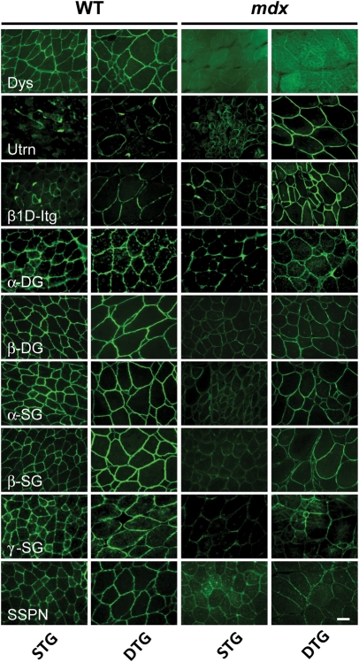
Broad sarcolemmal distribution of compensatory proteins upon Akt activation. Immunohistochemical analyses on transverse quadriceps sections in WT STG, WT DTG, mdx STG and mdx DTG mice. Sections were stained with antibodies to dystrophin (Dys), utrophin (Utrn), β1D integrin, alpha- and beta-dystroglycan (α-DG, β-DG), alpha-, beta- and gamma-sarcoglycan (α-SG, β-SG, γ-SG) and sarcospan (SSPN), and visualized using indirect immunofluorescence. Increased expression of the DGC and UGC in WT mice was observed upon constitutive activation of Akt1. Akt activation increased expression of only the UGC in mdx mice. An increase in utrophin levels was observed in mdx mice relative to levels in WT mice. In both WT mice and in mdx mice, Akt activation increased the expression of β1D integrin. Bar, 50 µm.
References
-
- Hoffman E.P., Brown R.H., Kunkel L.M. Dystrophin: the protein product of the Duchenne muscular dystrophy locus. Cell. 1987;51:919–928. - PubMed
-
- Ervasti J.M., Ohlendieck K., Kahl S.D., Gaver M.G., Campbell K.P. Deficiency of a glycoprotein component of the dystrophin complex in dystrophic muscle. Nature. 1990;345:315–319. - PubMed
-
- Durbeej M., Campbell K.P. Muscular dystrophies involving the dystrophin-glycoprotein complex: an overview of current mouse models. Curr. Opin. Genet. Dev. 2002;12:349–361. - PubMed
-
- Heydemann A., McNally E.M. Consequences of disrupting the dystrophin-sarcoglycan complex in cardiac and skeletal myopathy. Trends Cardiovasc. Med. 2007;17:55–59. - PubMed
Publication types
MeSH terms
Substances
Grants and funding
LinkOut - more resources
Full Text Sources
Other Literature Sources
Medical
Molecular Biology Databases
