. 2009 Jan 2;284(1):649-659.
doi: 10.1074/jbc.M806389200. Epub 2008 Nov 5.

Hey1 basic helix-loop-helix protein plays an important role in mediating BMP9-induced osteogenic differentiation of mesenchymal progenitor cells

Affiliations

Hey1 basic helix-loop-helix protein plays an important role in mediating BMP9-induced osteogenic differentiation of mesenchymal progenitor cells

Katie A Sharff et al. J Biol Chem. .

Abstract

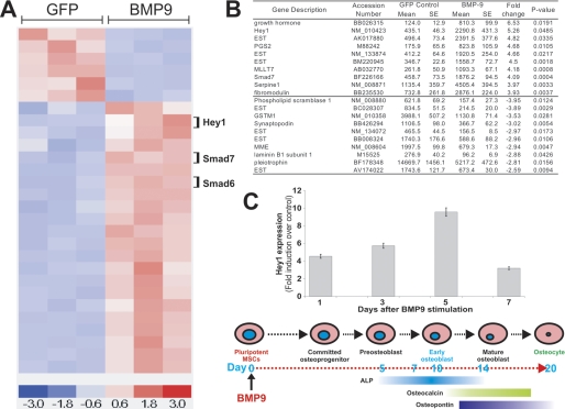
Pluripotent mesenchymal stem cells (MSCs) are bone marrow stromal progenitor cells that can differentiate into osteogenic, chondrogenic, adipogenic, and myogenic lineages. We previously demonstrated that bone morphogenetic protein (BMP) 9 is one of the most potent and yet least characterized BMPs that are able to induce osteogenic differentiation of MSCs both in vitro and in vivo. Here, we conducted gene expression-profiling analysis and identified that Hey1 of the hairy/Enhancer of split-related repressor protein basic helix-loop-helix family was among the most significantly up-regulated early targets in BMP9-stimulated MSCs. We demonstrated that Hey1 expression was up-regulated at the immediate early stage of BMP9-induced osteogenic differentiation. Chromatin immunoprecipitation analysis indicated that Hey1 may be a direct target of the BMP9-induced Smad signaling pathway. Silencing Hey1 expression diminished BMP9-induced osteogenic differentiation both in vitro and in vivo and led to chondrogenic differentiation. Likewise, constitutive Hey1 expression augmented BMP9-mediated bone matrix mineralization. Hey1 and Runx2 were shown to act synergistically in BMP9-induced osteogenic differentiation, and Runx2 expression significantly decreased in the absence of Hey1, suggesting that Runx2 may function downstream of Hey1. Accordingly, the defective osteogenic differentiation caused by Hey1 knockdown was rescued by exogenous Runx2 expression. Thus, our findings suggest that Hey1, through its interplay with Runx2, may play an important role in regulating BMP9-induced osteoblast lineage differentiation of MSCs.


Figures

FIGURE 1.
FIGURE 1.
Identification and verification of Hey1 as an early target of BMP9 signaling. A, dChip clustering analysis of the top 40 most significantly regulated genes by BMP9 in MSCs. B, list of the top 10 up-regulated and down-regulated target genes of BMP9 in MSCs. C, verification of BMP9-induced Hey1 expression in MSCs. Subconfluent C3H10T1/2 cells were infected with AdBMP9 or AdGFP. Total RNA was collected at the indicated times and subjected to qPCR analysis. The timeline of MSC differentiation leading to osteocytes is indicated at the bottom. All samples were normalized for glyceraldehyde-3-phosphate dehydrogenase expression. Reactions were done in duplicate. The fold induction was calculated by dividing the transcript level of BMP9-treated samples with that from GFP-treated samples.
FIGURE 2.
FIGURE 2.
ChIP analysis indicates that Hey1 is a direct target of BMP9-induced Smad signaling. A, schematic depiction of mouse Hey1 promoter region. The approximate locations of the three pairs of primers and their expected product sizes are indicated. B, subconfluent C3H10T1/2 cells were infected with AdGFP or AdBMP9 for 30 h. Cells were cross-linked. Genomic DNA was sonicated, following immunoprecipitation with anti-Smad4 or IgG. The retrieved genomic DNA was subjected to PCR using the three pairs of primers, PP-1, PP-2, and PP-3. The arrows indicate the locations of the expected products. C, control assays demonstrated that a similar amount of input materials was used for immunoprecipitation experiments. The arrows indicate the locations of the expected products. D, ChIP assays were carried out essentially the same as that described in B and C, except that Smad1/5/8 antibody was used for pulldown experiments. The arrows indicate the locations of the expected products. The ChIP analysis was performed in three independent experiments, and the representative results are shown.
FIGURE 3.
FIGURE 3.
Inhibition of BMP9-induced osteogenic differentiation by RNA interference-mediated knockdown of Hey1 gene expression. A, selection and verification of siRNAs targeting mouse Hey1. The target sites were subcloned and tested using the pSOS system (32). The resultant vectors were transfected into 293 cells, and knockdown of chimeric GFP/Hey1 expression was recorded 5 days after transfection. B, verification of Hey1 knockdown in the stable line C3H10-Hey1KD. Total RNA was collected from subconfluent stable and control lines, and subjected to qPCR analysis using primers corresponding to the 3′-untranslated repeat of mouse Hey1 (pUC19 as a DNA quantitation standard). All samples were normalized for glyceraldehyde-3-phosphate dehydrogenase expression. C and D, inhibition of ALP activity by Hey1 gene knockdown in MSCs. RNA interference-mediated knockdown of the Hey1 expression reduced BMP9-stimulated ALP activity as compared with the control line (C3H10T1/2) demonstrated by colorimetric assays at days 7 and 10 (C) and by histochemical staining at day 10 (D). Representative results of three independent experiments are shown.
FIGURE 4.
FIGURE 4.
Constitutive Hey1 expression enhances BMP9-induced osteogenic differentiation of MSCs. A, confirmation of Hey1 expression in the C3H10-Hey1 stable line. Total RNA was collected from subconfluent cells and subjected to qPCR analysis. All samples were normalized for glyceraldehyde-3-phosphate dehydrogenase. B and C, synergistic induction of BMP9-induced ALP activity by Hey1 overexpression. C3H10-Hey1 and C3H10T1/2 control lines were infected with AdGFP or AdBMP9. Alternatively, C3H10T1/2 cells were infected with AdGFP, AdBMP9, and/or AdHey1. ALP activity was determined at indicated time points using colorimetric assays (B) or histochemical staining (C). Representative results of three independent experiments are shown.
FIGURE 5.
FIGURE 5.
Critical role of Hey1 in BMP9-induced ectopic bone formation. C3H10-Hey1KD, C3H10-Hey1, and C3H10T1/2 control lines were transduced with AdBMP9 or AdGFP in vitro and implanted subcutaneously in athymic nude mice. At 6 weeks, animals were sacrificed and subjected to MicroCT imaging. A, MicroCT imaging analysis. Representative three-dimensional reconstructed images are shown. The injections sites are indicated by arrows. B, volumetric analysis of ectopic bone formation. Knockdown of Hey1 expression decreased ectopic bone formation (C3H10-Hey1KD = 0.27 mm3, C3H10T1/2 = 7.16 mm3, p = 0.02). Hey1 overexpression also reduced ectopic bone formation, although not statistically significant (C3H10-Hey1 = 3.5 mm3, C3H10T1/2 = 7.16 mm3, p = 0.08). C, histologic hematoxylin and eosin staining of retrieved samples. CC, chondrocyte; CM, chondroid matrix; MM, mineralized matrix; OB, osteoblast; OC, osteocyte; UD, undifferentiated MSCs. D, Trichrome (panels a and b) and Alcian Blue (panels c and d) staining of the samples retrieved AdBMP9 or AdBMP9 and Ad-simHey1 infected MEFs (3 weeks).
FIGURE 6.
FIGURE 6.
Inhibition of BMP9-mediated osteogenic differentiation due to Hey1 deficiency can be partially rescued by Runx2. A, Hey1 regulates expression of the late osteogenic marker. C3H10-Hey1KD and control lines were infected with AdBMP9 or AdGFP. Total RNA was collected at the indicated time points and subjected to qPCR analysis of osteocalcin (OC) expression. B, Runx2 and BMP9 act synergistically in the C3H10-Hey1KD line to increase osteogenic differentiation. Subconfluent C3H10-Hey1KD cells were infected with a low titer of AdBMP9 and increasing doses of AdRunx2. At indicated time points, cells were collected for colorimetric assays of ALP activity. C, Runx2 rescue on Hey1 knockdown was demonstrated in vivo by MicroCT imaging. D, Runx2 rescues the Hey1 knockdown phenotype. MicroCT imaging and volumetric data acquisition were carried out as described in Fig. 5B. E, histologic evidence of the Runx2 rescue effect on the Hey1KD MSCs. MM, mineralized matrix; OC, osteocyte; OB, osteoblast; UD, undifferentiated MSCs; magnification, 200×. F, regulation of Runx2 expression by Hey1. Total RNA was isolated from C3H10-Hey1KD and C3H10T1/2 control lines, and subjected to qPCR analysis for mouse Runx2 expression. See text for details.

References

    1. Pittenger, M. F., Mackay, A. M., Beck, S. C., Jaiswal, R. K., Douglas, R., Mosca, J. D., Moorman, M. A., Simonetti, D. W., Craig, S., and Marshak, D. R. (1999) Science 284 143-147 - PubMed
    1. Prockop, D. J. (1997) Science 276 71-74 - PubMed
    1. Aubin, J. E. (1998) J. Cell. Biochem. supplemental 31 73-82 - PubMed
    1. Ducy, P., Schinke, T., and Karsenty, G. (2000) Science 289 1501-1504 - PubMed
    1. Caplan, A. I., and Bruder, S. P. (2001) Trends Mol. Med. 7 259-264 - PubMed

Publication types

MeSH terms