. 2009 Jan;83(2):1126-34.
doi: 10.1128/JVI.01859-08. Epub 2008 Nov 5.

Characterization of large conformational changes and autoproteolysis in the maturation of a T=4 virus capsid

Affiliations

Characterization of large conformational changes and autoproteolysis in the maturation of a T=4 virus capsid

Tsutomu Matsui et al. J Virol. 2009 Jan.

Abstract

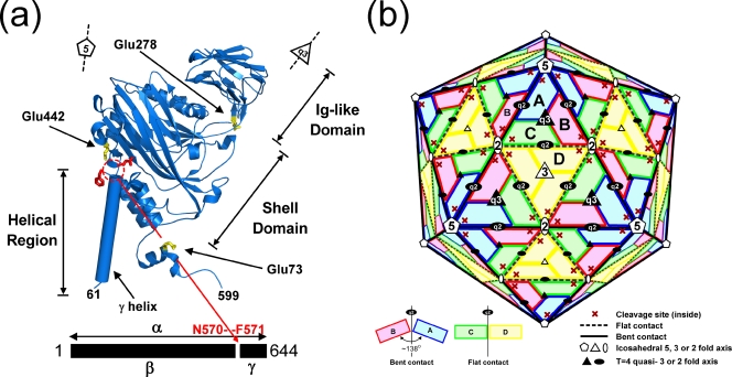
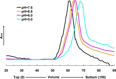
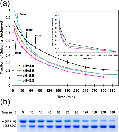
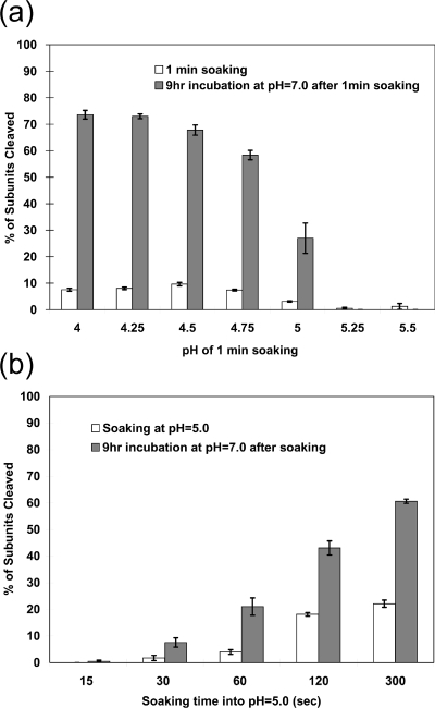
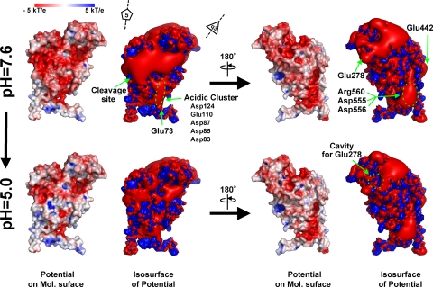
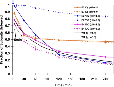
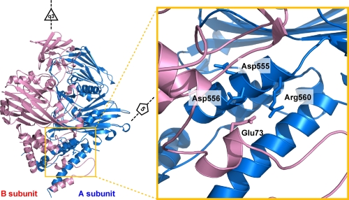
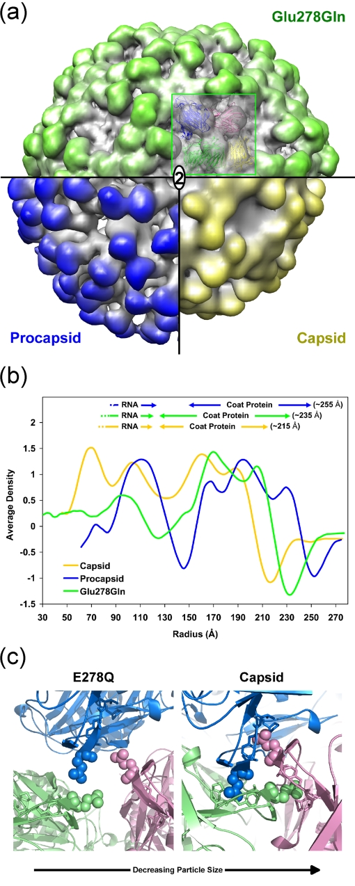
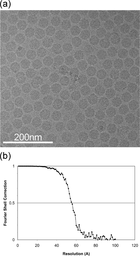
Nudaurelia capensis omega virus-like particles have been characterized as a 480-A procapsid and a 410-A capsid, both with T=4 quasisymmetry. Procapsids transition to capsids when pH is lowered from 7.6 to 5.0. Capsids undergo autoproteolysis at residue 570, generating the 74-residue C-terminal polypeptide that remains with the particle. Here we show that the particle size becomes smaller under conditions between pH 6.8 and 6.0 without activating cleavage and that the particle remains at an intermediate size when the pH is carefully maintained. At pH 5.8, cleavage is very slow, becoming detectable only after 9 h. The optimum pH for cleavage is 5.0 (half-life, approximately 30 min), with a significant reduction in the cleavage rate at pH values below 5. We also show that lowering the pH is required only to make the virus particles compact and to presumably form the active site for autoproteolysis but not for the chemistry of cleavage. The cleavage reaction proceeds at pH 7.0 after approximately 10% of the subunits cleave at pH 5.0. Employing the virion crystal structure for reference, we investigated the role of electrostatic repulsion of acidic residues in the pH-dependent large conformational changes. Three mutations of Glu to Gln that formed procapsids showed three different phenotypes on maturation. One, close to the threefold and quasithreefold symmetry axes and far from the cleavage site, did not mature at pH 5, and electron cryomicroscopy reconstruction showed that it was intermediate in size between those of the procapsid and capsid; one near the cleavage site exhibited a wild-type phenotype; and a third, far from the cleavage site, resulted in cleavage of 50% of the subunits after 4 h, suggesting quasiequivalent specificity of the mutation.


Figures

FIG. 1.
FIG. 1.
(a) The structure of the A subunit determined by the crystallographic analysis and refinement of NωV. The autoproteolytic cleavage occurs between Asn570 and Phe571 (shown as red stick side chains). The locations of three mutations discussed in the manuscript (Glu73, Glu278, and Glu442) are shown as yellow stick side chains. The structures of the B, C, and D subunits are closely similar to that of the A subunit, with some variations in the helical domain. (b) A schematic drawing of the T=4 subunit arrangement in the NωV particle. The quasiequivalent A, B, C, and D subunits are shown in blue, pink, green, and yellow, respectively. This color coding is maintained in all the figures that follow. Icosahedral and quasirotational axes are shown with white and black symbols, respectively. Note that the icosahedral twofold axes are quasisixfold axes in this surface lattice. Subunit contacts at the A/B interface (bent) have a dihedral angle of ∼138° (solid black line in the particle) and at the C/D interface (flat) have a dihedral angle of 180° (broken line).
FIG. 2.
FIG. 2.
The velocity sucrose gradient (10 to 30%) analysis of the cleavage-defective Asn570Thr mutant. The analysis was performed by placing particles initially at pH 7.6 onto gradients between pH 7.6 and 5.0. Centrifugation at 40,000 rpm was performed immediately and maintained for 1.25 h (corresponding to the incubation time at the designated pH). Peaks in the gradients at pH 7.6 and 5.0 correspond to the procapsid and capsid velocities, respectively. The sedimentation velocities at pH 6.8 and 6.0 indicate intermediate particle sizes. Between pH 6.0 and 5.0, it was impossible to detect differences in the sedimentation velocity of the particles even though, in the wild type, the proteolytic cleavage was not detected at pH 6.0 and proceeded relatively rapidly (see below) at pH 5. The latter observation indicates that there is a difference in the extent of the LCC at these two pH values; however, the experiment had insufficient resolution to detect this.
FIG. 3.
FIG. 3.
(a) Kinetics of cleavage measured for wild-type VLPs at different pH values. VLP procapsids were exposed to the designated pH, and aliquots were frozen in liquid nitrogen at the time points indicated. SDS gel analysis was then performed, and the fraction of subunits cleaved was determined by optical scanning of the gels to determine the amount of alpha and beta at the time point. The maximum initial rate was measured for particles incubated at pH 5. (b) A typical SDS-PAGE gel (wild-type NωV) under conditions of a time course incubation at pH 5.0.
FIG. 4.
FIG. 4.
Autoproteolysis occurs at neutral pH after activation at low pH. (a) The progress of the autoproteolytic cleavage at neutral pH values following activation at lower pH. The wild-type VLP procapsids were soaked in buffers between pH 4.00 and 5.50 for 1.0 min. The pH was then immediately shifted to pH 7.0, and the particles were incubated for 9.0 h. The fractions of subunits cleaved at 1 min and 9 h were determined as described for Fig. 3. (b) The progress of the autoproteolytic cleavage at neutral pH values following incubation at pH 5.0 for between 15 and 300 s. After being soaked at the indicated time points, the pH values were immediately changed to pH 7.0 and the mixture was incubated for 9 h. The chemistry of the reaction does not require low pH. The data indicate that ∼10% of the subunits must initially cleave at low pH for the reaction to proceed at pH 7.0, a result consistent with the observations of Canady et al. (7). We conclude that when fewer subunits cleave, the LCC is reversible. Panel b shows a relationship between the amount of initial cleavage and that seen after the reaction proceeds, suggesting that acidic residues with pKa values above 5.0 would play a key role in the LCC at low pH values.
FIG. 5.
FIG. 5.
Electrostatic potential of an isolated A subunit at pH 7.6 and 5.0; the molecular surface is colored by ±5-kT/e potentials calculated at the solvent-accessible surface and shown as blue and red gradations, respectively. Also, the ±3-kT/e potential isocontours are shown as blue and red surfaces, respectively. The view of both representations (left four panels) corresponds to Fig. 1a. The electrostatic potential was calculated for the monomeric protein, meaning that these potentials are not be influenced by the low-level dielectric environment or the charge distribution of neighboring subunits.
FIG. 6.
FIG. 6.
The cleavage assay of mutants at pH 5.0 and 4.5. The data for the wild type (Fig. 3a) are shown in black for comparison. An arrow at the top indicates the time point (2 min) at which Glu278Gln VLPs at pH 5.0 were frozen for cryoEM analysis.
FIG. 7.
FIG. 7.
A closeup view in the region near Glu73 (B subunit; shown in pink). Glu73 is located at the interface of subunits related by icosahedral and quasithreefold symmetry and occupies virtually identical positions in all four subunits. It forms a salt bridge with the neighboring Arg560 (A subunit; shown in blue) that is surrounded by negative charges (Arg560 and Asp556), as shown in Fig. 5. The phenotype of this mutation was unexpected, because only half the subunits cleave at pH 5.0 and 4.5, indicating an effect of quasiequivalence, and yet the residue displays high-fidelity quasiequivalence.
FIG. 8.
FIG. 8.
(a) A surface volume representation of the Glu278Gln mutant at 9.8-Å resolution (green), wild-type capsid at 25 Å resolution (yellow), and wild-type procapsid at 28-Å resolution (blue) (6). A pseudoatomic model of the mutant particle was generated by fitting the atomic coordinates, as a rigid body, to the observed density values. 2, twofold axis. (b) Comparison of the Glu278Gln mutant capsid, wild-type capsid, and wild-type procapsid (6) radially averaged electron density distributions. The averaged outer edge of the mutant corresponded to an intermediate size (∼235 Å) compared to those of the procapsid (∼255 Å) and capsid (∼215 Å). (c) A view of the quasithreefold axis near Glu278. At the left is a closeup view of the Gln278 pseudoatomic model described for panel a at pH 5.0. Glu278 is shown in the crystal structure of the capsid at the right. In the capsid form, Glu278 is inserted into a cavity formed in the neighboring subunit. The cavity consists of residues Ala276, Phe282, Val283, and Pro489 (side chains are shown as stick models). These hydrophobic residues are surrounded by negative charges at pH 7.6; however, the charge is reduced at pH 5.0 (Fig. 5). The LCC in the Glu278Gln particle was inhibited by this substitution, indicating that the self-repulsion between symmetry-related Glu278 residues plays a crucial role in the cavity insertion.
FIG. 9.
FIG. 9.
(a) A typical micrograph of the Glu278Gln mutant particles flash frozen and imaged at ×80,000 magnification. (b) Resolution determination of the reconstruction based on the Fourier shell correlation method. The particle images used for the reconstruction refinement were divided into equally sized data sets, and two new reconstructions were calculated. The correlation coefficients of the two resulting Fourier shell reconstructions were then computed to estimate the resolution. The 0.5 Fourier shell correlation criterion was 9.8 Å.

References

    1. Agrawal, D. K., and J. E. Johnson. 1995. Assembly of the T = 4 Nudaurelia capensis omega virus capsid protein, post-translational cleavage, and specific encapsidation of its mRNA in a baculovirus expression system. Virology 20789-97. - PubMed
    1. Agrawal, D. K., and J. E. Johnson. 1992. Sequence and analysis of the capsid protein of Nudaurelia capensis omega virus, an insect virus with T = 4 icosahedral symmetry. Virology 190806-814. - PubMed
    1. Baker, N. A., D. Sept, S. Joseph, M. J. Holst, and J. A. McCammon. 2001. Electrostatics of nanosystems: application to microtubules and the ribosome. Proc. Natl. Acad. Sci. USA 9810037-10041. - PMC - PubMed
    1. Brooks, E. M., K. H. Gordon, S. J. Dorrian, E. R. Hines, and T. N. Hanzlik. 2002. Infection of its lepidopteran host by the Helicoverpa armigera stunt virus (Tetraviridae). J. Invertebr. Pathol. 8097-111. - PubMed
    1. Brünger, A. T., P. D. Adams, G. M. Clore, W. L. DeLano, P. Gros, R. W. Grosse-Kunstleve, J. S. Jiang, J. Kuszewski, M. Nilges, N. S. Pannu, R. J. Read, L. M. Rice, T. Simonson, and G. L. Warren. 1998. Crystallography & NMR system: a new software suite for macromolecular structure determination. Acta Crystallogr. D Biol. Crystallogr. 54905-921. - PubMed

Publication types

LinkOut - more resources