. 2009 Jan;83(2):1093-104.
doi: 10.1128/JVI.01633-08. Epub 2008 Nov 5.

Human immunodeficiency virus type 1 Nef incorporation into virions does not increase infectivity

Affiliations

Human immunodeficiency virus type 1 Nef incorporation into virions does not increase infectivity

Nadine Laguette et al. J Virol. 2009 Jan.

Abstract

The viral protein Nef contributes to the optimal infectivity of human and simian immunodeficiency viruses. The requirement for Nef during viral biogenesis particles suggests that Nef might play a role in this process. Alternatively, because Nef is incorporated into viruses, it might play a role when progeny virions reach target cells. We challenged these hypotheses by manipulating the amounts of Nef incorporated in viruses while keeping its expression level constant in producer cells. This was achieved by forcing the incorporation of Nef into viral particles by fusing a Vpr sequence to the C-terminal end of Nef. A cleavage site for the viral protease was introduced between Nef and Vpr to allow the release of Nef fragments from the fusion protein during virus maturation. We show that the resulting Nef-CS-Vpr fusion partially retains the ability of Nef to downregulate cell surface CD4 and that high amounts of Nef-CS-Vpr are incorporated into viral particles compared with what is seen for wild-type Nef. The fusion protein is processed during virion maturation and releases Nef fragments similar to those found in viruses produced in the presence of wild-type Nef. Unlike viruses produced in the presence of wild-type Nef, viruses produced in the presence of Nef-CS-Vpr do not have an increase in infectivity and are as poorly infectious as viruses produced in the absence of Nef. These findings demonstrate that the presence of Nef in viral particles is not sufficient to increase human immunodeficiency virus type 1 infectivity and suggest that Nef plays a role during the biogenesis of viral particles.


Figures
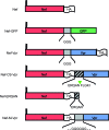
FIG. 1.
FIG. 1.
Nef-based constructs used in this study. The various domains of the chimeric proteins are illustrated. Except for Nef-GFP, all constructs are HA tagged to facilitate the immunodetection in Western blotting experiments. The HA tag was added to the C-terminal ends of the proteins in order to preserve the myristoylation site of Nef. In the case of Nef-CS-Vpr, an HIV-1 protease recognition motif corresponding to the NC-p1 junction in the Gag-Pol precursor was inserted between Nef and Vpr. The HA tag was moved between Nef and this motif in order to visualize Nef fragments generated from the cleavage of the fusion protein in the virus. A similar construct was also generated that contains a glycine stretch in place of the protease recognition motif. Because the processing of Nef-CS-Vpr is supposed to release Nef fragments with the ERQAN sequence C terminal to the HA tag, a Nef-ERQAN was also generated and analyzed in incorporation and infectivity experiments.
FIG. 2.
FIG. 2.
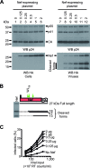
Expression of Nef during HIV-1 production: impact on viral infectivity. GFP reporter viruses were produced in 293T cells in the presence of increasing amounts of a Nef-coding plasmid (0.125 to 2 μg/T75 flasks). (A) Producer cells were solubilized (left) and viruses were pelleted from the cell culture supernatant by ultracentrifugation (right). Samples were resolved by SDS-PAGE and visualized by Western blotting (WB) followed by immunodetection with p24 (top)- and HA (bottom)-specific antibodies. (B) Molecular species derived from the processing of Nef in mature viruses. The lane showing the incorporation of Nef into viral particles (panel A, lane 10) is shown next to a schematic representation of Nef processed by HIV-1 protease. The position of the processing sites (green arrows) was determined according to published data and the measurement of the mobility of the anti-HA reactive proteins in the gel. (C) Viral infectivity was assayed with HeLa CD4 cells. HeLa CD4 cells were incubated with increasing amounts of viruses (50 × 103 to 400 × 103 RT cpm/ml) produced in the presence of the indicated amounts of Nef-coding plasmid, and the percentages of GFP-positive cells were measured by fluorescence-activated cell sorting 60 h postinfection.
FIG. 3.
FIG. 3.
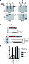
Characterization of viruses produced in the presence of Nef, Nef-Vpr, and Vpr. 293T cells were cotransfected with Gag-Pol in combination with either Nef, Nef-Vpr, or Vpr expression plasmids as indicated, along with the plasmids required for the production of infectious particles. (A) Cells and viruses were analyzed as described in the Fig. 2A legend. (B) The processing of the Nef-Vpr fusion protein (bottom) was analyzed as described in the Fig. 2B legend and compared to that of WT Nef (top). Thick arrows indicate Nef-WT cleavage sites; thin arrows indicate three additional sites where Nef-Vpr also appears to be susceptible to the viral protease. (C) HXBc2 Env- or VSV-G-pseudotyped viruses were produced in the absence or in the presence of WT Nef or the Nef-Vpr fusion protein. Viral infectivity was measured as described in the Fig. 2C legend and normalized to that of viruses produced in the presence of Nef Xho.
FIG. 4.
FIG. 4.
CD4 downregulation activity and subcellular localization of Nef-Vpr. (A) Cell surface CD4 expression was analyzed with cells coexpressing CD4 or CD4ΔCT along with Nef variants or Vpr as indicated. Cells were also cotransfected with a plasmid encoding GFP to gate the analysis of cell surface CD4 expression on transfected cells. Dot plots of cells stained with a CD4-specific antibody are shown. (B) Quantification of the CD4 downregulation activity of Nef variants measured as described for panel A. (C) Cellular distribution of Nef, Nef-Vpr, and Vpr. HeLa cells were grown on coverslips and transfected with the indicated HA-tagged constructs. Twenty-four hours posttransfection, cells were permeabilized and stained with an HA-specific antibody and an Alexa488-coupled secondary antibody. Coverslips were transferred to slides in mounting medium supplemented with DAPI and samples were analyzed by immunofluorescence microscopy followed by deconvolution as described in Materials and Methods.
FIG. 5.
FIG. 5.
Differential consequences of the addition of GFP or Vpr to the C-terminal end of Nef. (A) Viruses were produced in the presence of WT Nef (left) or Nef-GFP (right). Cells and viruses were analyzed as described in the Fig. 2A legend. Nef-GFP was detected with GFP-specific antibodies. (B) Viral infectivities of viruses produced in the presence of the indicated variants of Nef were analyzed as described in the Fig. 2C legend. (C) Viruses were produced in the presence of the indicated combination of Nef, Nef-Vpr, or Vpr. Cells and viruses were analyzed as described in the Fig. 2A legend. Arrowheads point out the 27- and the 18-kDa forms of Nef. (D) Viral infectivities of viruses produced in the presence of the indicated combinations of Nef, Nef-Vpr, and/or Vpr were measured as indicated in the Fig. 2C legend and normalized to that of viruses produced in the presence of Nef Xho.
FIG. 6.
FIG. 6.
Characterization of a Nef-Vpr fusion protein containing a protease specific cleavage site between Nef and Vpr. (A) Viruses were produced in the presence of WT Nef, Nef-M-Vpr, or Nef-CS-Vpr as indicated. Cells (left) and viruses (middle) were analyzed as described in the Fig. 2A legend. Viral core purification (right) was performed by pelleting cell culture supernatant through a thin detergent layer as indicated in Materials and Methods. Viral matrix and capsid were detected with p17- and p24-specific antibodies, respectively. Film obtained with a short exposure time (30 s) is shown in order to better distinguish Nef-Vpr-derived fragments in whole viruses and cores. This explains the weak signal obtained with Nef, since Nef-Vpr fusion proteins are better incorporated than Nef. (B) The processing of the Nef-M-Vpr (top) and Nef-CS-Vpr (bottom) fusion proteins was analyzed as described in the Fig. 2B legend. For Nef lanes, film obtained with a longer exposure time was used (5 min). The arrowhead indicates cleavage of the Nef-CS-Vpr fusion protein at the HIV-1 protease-specific cleavage site (CS) introduced between Nef and VPR. The ability of the indicated constructs to increase HIV-1 infectivity (C) and to downregulate cell surface CD4 (D) was analyzed as described in the Fig. 2 and 4 legends, respectively.
FIG. 7.
FIG. 7.
Characterization of the Nef-ERQAN fusion protein. (A) Viruses were produced in the presence of the indicated combination of Nef, Nef-ERQAN, and Nef-CS-Vpr. Cells and viruses were analyzed as described in the Fig. 2A legend. The amounts of Nef-CS-Vpr-coding plasmid cotransfected with Nef and Nef-ERQAN were adjusted to achieve incorporation of similar amounts of Nef-CS-Vpr and Nef in viral particles. (B) Viral infectivities of viruses produced in the presence of the indicated combinations of Nef, Nef-ERQAN, and Nef-CS-Vpr were measured as indicated in the Fig. 2C legend and normalized to that of viruses produced in the presence of Nef Xho. * indicates a product cleaved by the viral protease.

Similar articles

Cited by

References

    1. Accola, M. A., A. A. Bukovsky, M. S. Jones, and H. G. Gottlinger. 1999. A conserved dileucine-containing motif in p6gag governs the particle association of Vpx and Vpr of simian immunodeficiency viruses SIVmac and SIVagm. J. Virol. 739992-9999. - PMC - PubMed
    1. Accola, M. A., A. Ohagen, and H. G. Gottlinger. 2000. Isolation of human immunodeficiency virus type 1 cores: retention of Vpr in the absence of p6gag. J. Virol. 746198-6202. - PMC - PubMed
    1. Ahmad, N., and S. Venkatesan. 1988. Nef protein of HIV-1 is a transcriptional repressor of HIV-1 LTR. Science 2411481-1485. - PubMed
    1. Aiken, C., and D. Trono. 1995. Nef stimulates human immunodeficiency virus type 1 proviral DNA synthesis. J. Virol. 695048-5056. - PMC - PubMed
    1. Aiken, C. 1997. Pseudotyping human immunodeficiency virus type 1 (HIV-1) by the glycoprotein of vesicular stomatitis virus targets HIV-1 entry to an endocytic pathway and suppresses both the requirement for Nef and the sensitivity to cyclosporin A. J. Virol. 715871-5877. - PMC - PubMed

Publication types

Substances