. 2009 Jan;83(2):870-83.
doi: 10.1128/JVI.01088-08. Epub 2008 Nov 5.

Identification of the LWYIK motif located in the human immunodeficiency virus type 1 transmembrane gp41 protein as a distinct determinant for viral infection

Affiliations

Identification of the LWYIK motif located in the human immunodeficiency virus type 1 transmembrane gp41 protein as a distinct determinant for viral infection

Steve S-L Chen et al. J Virol. 2009 Jan.

Abstract

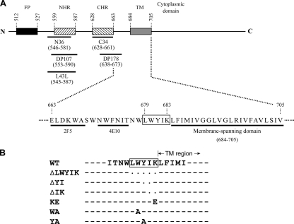
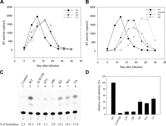
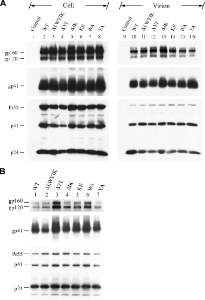
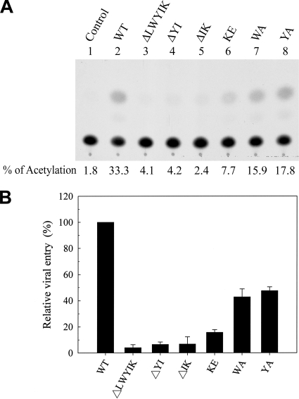
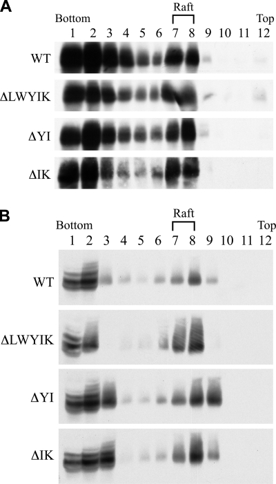
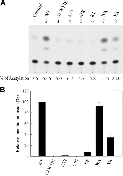
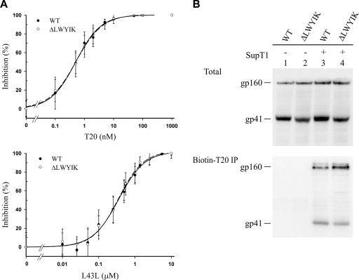
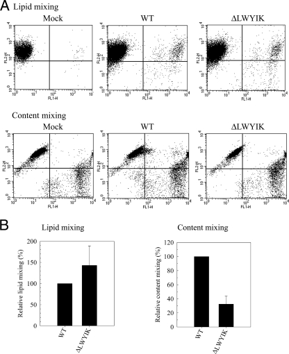
The highly conserved LWYIK motif located immediately proximal to the membrane-spanning domain of the gp41 transmembrane protein of human immunodeficiency virus type 1 has been proposed as being important for the surface envelope (Env) glycoprotein's association with lipid rafts and gp41-mediated membrane fusion. Here we employed substitution and deletion mutagenesis to understand the role of this motif in the virus life cycle. None of the mutants examined affected the synthesis, precursor processing, CD4 binding, oligomerization, or cell surface expression of the Env, nor did they alter Env incorporation into the virus. All of the mutants, particularly the DeltaYI, DeltaIK, and DeltaLWYIK mutants, in which the indicated residues were deleted, exhibited greatly reduced one-cycle viral replication and the Env trans-complementation ability. All of these deletion mutant proteins were still localized in the lipid rafts. With the exception of the Trp-to-Ala (WA) mutant, which exhibited reduced viral infectivity albeit with normal membrane fusion, all mutants displayed loss of some or almost all of the membrane fusion ability. Although these deletion mutants partially inhibited in trans wild-type (WT) Env-mediated fusion, they were more effective in dominantly interfering with WT Env-mediated viral entry when coexpressed with the WT Env, implying a role of this motif in postfusion events as well. Both T20 and L43L peptides derived from the two gp41 extracellular C- and N-terminal alpha-helical heptad repeats, respectively, inhibited WT and DeltaLWYIK Env-mediated viral entry with comparable efficacies. Biotin-tagged T20 effectively captured both the fusion-active, prehairpin intermediates of WT and mutant gp41 upon CD4 activation. Env without the deletion of the LWYIK motif still effectively mediated lipid mixing but inhibited content mixing. Our study demonstrates that the immediate membrane-proximal LWYIK motif acts as a unique and distinct determinant located in the gp41 C-terminal ectodomain by promoting enlargement of fusion pores and postfusion activities.


Figures

FIG. 1.
FIG. 1.
(A) Schematic representation of gp41. The rectangles marked FP, NHR, CHR, and TM indicate the locations of the fusion peptide, N-terminal HR sequence, C-terminal HR sequence, and TM domain, respectively. Residues 663 to 683 of the extracellular domain, which constitute the Trp-rich region, and the TM domain are shown in the single-amino-acid code. The immediate membrane-proximal LWYIK motif, located at residues 679 to 683, is boxed. The epitopes for the 2F5 and 4E10 MAbs and the locations of the inhibitor peptides are underlined with dark lines. (B) Construction of the LWYIK motif mutant proviruses. Site-specific, oligonucleotide-directed mutagenesis was performed based on the infectious pHXB2RU3 provirus to generate proviral variants with mutations in the LWYIK motif as indicated. Dashes and dots indicate that the residue in that position of the mutant proviruses is identical to or absent from that of the WT provirus, respectively.
FIG. 2.
FIG. 2.
Effects of mutations in the LWYIK motif on viral infectivity. (A and B) Replication kinetics of viruses. Cell-free WT and mutant viruses containing equal amounts of RT activity were used to challenge CEM-SS cells, and virus production in the culture media as measured by RT activity was monitored after infection. (C and D) One-cycle viral replication assay. H938 cells were infected with RT-normalized WT or mutant viruses. Two days after infection, cell lysates were prepared, and CAT activity was determined. A typical CAT activity analysis is shown in panel C. The background CAT level detected in mock-infected cells (control) was subtracted from the CAT activity of the WT or mutant virus infection. The relative infectivities of mutant viruses are expressed as percentages of that of the WT virus. The results from four independent experiments (mean ± standard deviation) are shown in panel D.
FIG. 3.
FIG. 3.
Assessment of viral protein expression. (A) HeLa cells were mock transfected (control) or transfected with the WT or mutant proviruses, and equal volumes of cell and virion lysates were subjected to SDS-PAGE followed by Western blotting analysis using the 902, Chessie 8, and 183 MAbs. (B) Cell-free culture supernatants obtained from acutely infected CEM-SS cells at the peak of viral replication were concentrated by ultracentrifugation and reconstituted with RPMI containing 1% FBS, and p24 levels were determined. WT and mutant virions containing equal amounts of p24 were resolved by SDS-PAGE followed by Western blotting using the 902, Chessie 8, and 183 MAbs.
FIG. 4.
FIG. 4.
Characterization of LWYIK motif mutant Env proteins. (A) Cell surface expression. HeLa cells mock transfected (control) or transfected with the WT or mutant proviruses were metabolically labeled with [35S]methionine and surface biotinylated with sulfo-N-hydroxysuccinimide-biotin. Levels of Env proteins expressed in cells (top panel) or on the cell surface (bottom panel) were assessed as described in Materials and Methods. (B) CD4-binding ability. HeLa cells transfected with pSVE7puro(ΔKS) (control), WT, or mutant pSVE7puro plasmids in the presence of pIIIextat were metabolically labeled with [35S]methionine. Levels of Env proteins expressed in cells (top panel) and the CD4-binding ability of Env proteins (bottom panel) were determined. (C) Oligomerization ability. HeLa cells were transfected with pSVE7puro(ΔKS) (control), WT, or TC mutant pSVE7puro plasmid or cotransfected with the TC plasmid along with the WT or LWYIK motif plasmids in the presence of pIIIextat. Levels of Env proteins expressed in cells (top panel) and the self-assembly abilities of the Env proteins (bottom panel) were determined.
FIG. 5.
FIG. 5.
Env trans-complementation assay of mutant Env proteins. Env pseudotypes produced from 293T cells cotransfected with pHXB2ΔBglCAT and each of the ΔKS (control), WT, or mutant Env plasmids were normalized for RT activity before being used to challenge CEM-SS cells. CAT activity was measured 3 days after virus infection. A representative result is shown in panel A. The background CAT level detected in the defective virus produced from ΔKS pSVE7puro cotransfection was subtracted from the CAT activity of the WT or mutant pseudotypes. The relative viral entry ability mediated by mutant proteins is expressed as a percentage of that mediated by the WT Env. The results from three independent experiments are shown as the mean ± standard deviation in panel B.
FIG. 6.
FIG. 6.
Effects of deletions in the LWYIK motif on Env localization in lipid rafts. (A) 293T cells were transfected with 10 μg each of the WT and mutant proviruses, and transfected cells were extracted with 1% Triton X-100 at 4°C. The lysates were subjected to lipid raft membrane flotation assay followed by Western blotting using the Chessie 8 MAb. The distribution profile of gp41 is shown. (B) CEM-SS cells acutely infected with WT or mutant viruses were collected at or near the peak of virus infection, extracted with 1% cold Triton X-100, and then subjected to a lipid raft fractionation analysis as described for panel A.
FIG. 7.
FIG. 7.
Effects of mutations in the LWYIK motif on the Env membrane fusion ability. 293T cells were cotransfected with pIIIextat along with the ΔKS (control), WT, or mutant pSVE7puro plasmids as indicated and cocultured with 106 H938 cells, and CAT activity was assessed. A representative result is shown in panel A. The background CAT level detected in the absence of Env (control) was subtracted from the CAT activity detected in the presence of the WT or mutant proteins. The relative membrane fusion activity of the mutant proteins is expressed as a percentage of that of the WT Env. The results from three independent experiments (mean ± standard deviation) are shown in panel B.
FIG. 8.
FIG. 8.
Ability of deletion mutants to dominantly interfere with WT Env-mediated membrane fusion and viral entry. (A) 293T cells were cotransfected with 1.5 μg of pIIIextat together with 10 μg of pSVE7puro(ΔKS) (control) (lane 1), 5 μg each of the WT and ΔKS plasmids (lane 2), or 5 μg each of the WT and mutant plasmids (lanes 3 to 6). Transfected cells were cocultured with H938 cells, and CAT activity was assayed. (B) 293T cells were cotransfected with 7.5 μg of pHXBΔBglCAT and 10 μg of pSVE7puro(ΔKS) (control) (lane 1), 5 μg each of the WT and ΔKS plasmids (lane 2), or 5 μg each of the WT and mutant pSVE7puro plasmids (lanes 3 to 6). Cell-free viruses normalized by RT activity from each transfection were used to challenge 106 CEM-SS cells, and CAT activity was assayed. In each case, a representative CAT assay is shown in the top panel, whereas the results from four (for membrane fusion) and three (for viral entry) independent experiments (mean ± standard deviation) are shown in the bottom panel.
FIG. 9.
FIG. 9.
Sensitivities of WT and ΔLWYIK mutant viruses to CHR and NHR peptides. (A) Inhibition of WT and ΔLWYIK mutant Env pseudotypes to L43L and T20 peptides. Replication-incompetent NL4-3RELuc viruses pseudotyped with WT or ΔLWYIK mutant proteins were incubated with T20 or L43L peptide at the concentrations indicated, and cell lysates were assayed for luciferase activity. Ten times more ΔLWYIK mutant pseudotyped virus, in terms of RT activity, than the WT pseudotype was examined. The percent inhibition of WT and mutant pseudotypes at each peptide concentration was calculated as described in Materials and Methods from at least five independent analyses, and the averages with standard deviations are shown. (B) 293T cells transiently expressing WT or ΔLWYIK mutant Env were incubated with biotinylated T20 in the presence or absence of SupT1 cells. The cell lysates were directly analyzed by Western blotting (top panel) or subjected to coprecipitation with neutravidin beads prior to Western blotting with Chessie 8 MAb (bottom panel).
FIG. 10.
FIG. 10.
Fluorescent probe exchange assays. (A) HeLa cells transfected with pCAGGS-based WT or ΔLWYIK mutant plasmids and labeled with DiO (green) were cocultured with CEM-SS cells labeled with DiI (red) as described in Materials and Methods. CEM-SS cells stained with both lipophilic fluorescent dyes were quantified by flow cytometry, and a representative two-color flow cytometic analysis is shown in the top panel. In addition, WT or ΔLWYIK mutant Env-expressing HeLa cells labeled with calcein-AM (green) and CEM-SS cells labeled with CMTMR (red) were cocultured, and CEM-SS cells stained with both cytosolic probes were quantified by flow cytometry; a representative result is shown in the bottom panel. (B) The background percentage of lipophilic dye transfer detected in pCAGGS vector transfection (control) was subtracted from that detected in WT or ΔLWYIK Env plasmid transfection, and the degree of lipid mixing of the ΔLWYIK mutant is expressed as a percentage of that of the WT Env. Results from three independent experiments (mean ± standard deviation) are shown (left panel). Also, the degree of content mixing of the ΔLWYIK mutant was quantified from four separate analyses and is expressed as a percentage of that of the WT Env (right panel).

References

    1. Anderson, R. G. 1998. The caveolae membrane system. Annu. Rev. Biochem. 67199-225. - PubMed
    1. Bellamy-McIntyre, A. K., C. S. Lay, S. Baar, A. L. Maerz, G. H. Talbo, H. E. Drummer, and P. Poumbourios. 2007. Functional links between the fusion peptide-proximal polar segment and membrane-proximal region of human immunodeficiency virus gp41 in distinct phases of membrane fusion. J. Biol. Chem. 28223104-23116. - PubMed
    1. Berman, P. W., W. M. Nunes, and O. K. Haffar. 1988. Expression of membrane-associated and secreted variants of gp160 of human immunodeficiency virus type 1 in vitro and in continuous cell lines. J. Virol. 623135-3142. - PMC - PubMed
    1. Bhattacharya, J., P. J. Peters, and P. R. Clapham. 2004. Human immunodeficiency virus type 1 envelope glycoproteins that lack cytoplasmic domain cysteines: impact on association with membrane lipid rafts and incorporation onto budding virus particles. J. Virol. 785500-5506. - PMC - PubMed
    1. Brown, D. A., and J. K. Rose. 1992. Sorting of GPI-anchored proteins to glycolipid-enriched membrane subdomains during transport to the apical cell surface. Cell 68533-544. - PubMed

Publication types

Substances