. 2008 Nov 5;28(45):11445-53.
doi: 10.1523/JNEUROSCI.1972-08.2008.

Impact of apolipoprotein E (ApoE) polymorphism on brain ApoE levels

Affiliations

Impact of apolipoprotein E (ApoE) polymorphism on brain ApoE levels

David R Riddell et al. J Neurosci. .

Abstract

Inheritance of the apoE4 allele (epsilon4) increases the risk of developing Alzheimer's disease; however, the mechanisms underlying this association remain elusive. Recent data suggest that inheritance of epsilon4 may lead to reduced apoE protein levels in the CNS. We therefore examined apoE protein levels in the brains, CSF and plasma of epsilon2/2, epsilon3/3, and epsilon4/4 targeted replacement mice. These apoE mice showed a genotype-dependent decrease in apoE levels; epsilon2/2 >epsilon3/3 >epsilon4/4. Next, we sought to examine the relative contributions of apoE4 and apoE3 in the epsilon3/4 mouse brains. ApoE4 represented 30-40% of the total apoE. Moreover, the absolute amount of apoE3 per allele was similar between epsilon3/3 and epsilon3/4 mice, implying that the reduced levels of total apoE in epsilon3/4 mice can be explained by the reduction in apoE4 levels. In culture medium from epsilon3/4 human astrocytoma or epsilon3/3, epsilon4/4 and epsilon3/4 primary astrocytes, apoE4 levels were consistently lower than apoE3. Secreted cholesterol levels were also lower from epsilon4/4 astrocytes. Pulse-chase experiments showed an enhanced degradation and reduced half-life of newly synthesized apoE4 compared with apoE3. Together, these data suggest that astrocytes preferentially degrade apoE4, leading to reduced apoE4 secretion and ultimately to reduced brain apoE levels. Moreover, the genotype-dependent decrease in CNS apoE levels, mirror the relative risk of developing AD, and suggest that low levels of total apoE exhibited by epsilon4 carriers may directly contribute to the disease progression, perhaps by reducing the capacity of apoE to promote synaptic repair and/or Abeta clearance.


Figures

Figure 1.
Figure 1.
Development of an apoE4-specific immunoassay. a, Western blotting of 1 μg/lane of recombinant apoE2, apoE3 and apoE4 standards using either an apoE4 selective or a nonselective pan apoE antibody. b, c, ApoE2 (▵), apoE3 (■) and apoE4 (○) standards were assayed using MSD-based homogenous immunoassays, as outlined in Materials and Methods, incorporating either the pan-apoE (b) or apoE4-specific antibodies (c). Results are expressed as MSD electrochemiluminescence (ECL) counts and each data point represents the mean ± SEM of three independent experiments with triplicates. d, Cortices from 20-week-old apoE-targeted replacement mice were detergent extracted, and apoE was measured in the tissue homogenates using either the pan-apoE immunoassay (total apoE) or the apoE4 selective assay and normalized to total protein levels as outlined in Materials and Methods (n = 4–14 animals per group; mean ± SEM).
Figure 2.
Figure 2.
ApoE-targeted replacement mice have a genotype-dependent decrease in brain, CSF and plasma apoE levels. a, Frontal cortex and hippocampus from 20-week-old apoE-targeted replacement mice were detergent extracted and total apoE was measured in the tissue homogenates using the pan-apoE immunoassay and normalized to total protein levels as outlined in Materials and Methods (n = 22 animals per group; mean ± SEM). b, Ten micrograms of cortical or hippocampal homogenates (from ε2/2, ε3/3 or ε4/4) were subjected to Western blotting using a polyclonal anti-pan-apoE antibody. Anti-α-tubulin was used as a loading control. c, mRNA expression levels of hApoE in the frontal cortex and hippocampus of 20-week-old targeted replacement mice were analyzed by quantitative real-time PCR. RNA amounts were normalized with 18 s (n = 4–10 animals per group; mean ± SEM). d, e, Plasma (d) or CSF (e) from 20-week-old apoE-targeted replacement mice were detergent extracted, and total apoE was measured using the pan-apoE immunoassay as outlined in Materials and Methods (n = 20 animals per group; mean ± SEM; **p < 0.01, ***p < 0.001, NS, nonsignificant).
Figure 3.
Figure 3.
Heterozygous ε3/4-targeted replacement mice have reduced levels of brain, CSF and plasma apoE4 relative to apoE3. a, Frontal cortex and hippocampus from 12-week-old apoE-targeted replacement mice were detergent extracted and total apoE was measured in the tissue homogenates using the pan-apoE immunoassay and normalized to total protein levels as outlined in Materials and Methods (n = 14–22 animals per group; mean ± SEM). b, Frontal cortex, hippocampus, CSF and plasma from 12-week-old ε3/4-targeted replacement mice were detergent extracted and total apoE and apoE4 protein levels were measured in the tissue homogenates using the pan- and apoE4-selective immunoassays, respectively. ApoE levels were normalized to total protein as outlined in Materials and Methods and the percentage of total apoE calculated for apoE3 and apoE4. (n = 10–22 animals per group; mean ± SEM; **p < 0.01, ***p < 0.001). c, d, The amount of apoE3 or apoE4 per allele was calculated for frontal cortex (c) and hippocampus (d). The amount of apoE3 per allele is similar between ε3/3 and ε3/4 mice.
Figure 4.
Figure 4.
Identification of human astrocytomas heterozygous for the ε4 allele. a, ApoE genotyping of human astrocytoma cell lines (CCF-STTG, U87 and U118) was performed by amplifying a 244-bp PCR product from exon IV and restriction isotyping with HhaI (Hixson and Vernier, 1990). Each genotype gives a specific combination of HhaI fragment sizes: ε2/2, 91 and 83 bp; ε3/3, 91 and 48 bp; ε4/4, 72 and 48 bp and a mixed genotype: ε2/3, 91, 83, and 48 bp; ε3/4, 91, 72 and 48 bp; ε2/4, 91, 83, 72 and 48 bp. CCF-STTG1 cells were genotyped as ε3/4, whereas U118 cells were ε2/4. b, DNA sequencing of the 244 bp PCR product from CCF-STTG1 cells verified that they exhibited a mixed ε3/4 genotype at codon 112. c, Conditioned media from CCF-STTG1 cells was analyzed by Western blotting using an apoE4 selective antibody (as outlined in Materials and Methods). Purified recombinant apoE3 and apoE4 were used as standards.
Figure 5.
Figure 5.
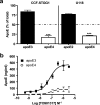
Reduced apoE4 levels in conditioned media from heterozygous human astrocytomas. a, CCF-STTG1 cells were cultured in serum-free media for 48 h. The media was removed, apoE-containing lipoproteins isolated by flotation ultracentrifugation (density <1.21 g/ml) and assayed for total apoE and apoE4 protein levels using the MSD immunoassays as outlined in Materials and Methods (n = 3, mean ± SEM, ***p < 0.001). Because U118 have very low basal levels of apoE secretion, U118 cells (ε2/4) were cultured in serum-free media containing 1 μm T0901317 for 48 h. The media was removed, concentrated 10-fold and assayed for total apoE and apoE4 protein levels using the MSD immunoassays as outlined in Materials and Methods (n = 3, mean ± SEM, ***p < 0.001). b, Dose-dependent upregulation of apoE3 and apoE4 by LXR agonist T0901317 in CCF-STTG1 cells. Cells were treated with T0901317 at the indicated concentrations for 48 h. Conditioned media were subjected to total apoE and apoE4 selective immunoassays as described in Materials and Methods. ApoE3 levels were calculated by subtracting apoE4 from total apoE levels.
Figure 6.
Figure 6.
Astrocytic apoE4 shows enhanced degradation and reduced secretion relative to apoE3. Primary astrocytes from apoE3/3 and apoE4/4 heterozygous mice were cultured 24 h in serum-free media, washed and incubated in methionine-free DMEM with 200 μCi/ml [35S]methionine for 3 h. Cells were then washed and chased with serum-free media containing unlabeled methionine. At indicated times, 35S-labeled apoE was immunoprecipitated from media and cell lysates, separated and detected by SDS-PAGE and phosphorimaging of a single 34 kDa band, as described in Materials and Methods. a–c, Cell-associated [35S]-apoE (a), secreted [35S]-apoE (b), and net degradation of [35S]-apoE (c), calculated by subtracting residual [35S]-apoE in cell and media from [35S]-apoE in cells at time 0 (T0), were measured. d, The effect of apoE genotype on the relative contribution of cell association, degradation and secretion of apoE at the 300 min time point. All data are percentages of total [35S]-apoE at T0.

References

    1. Beffert U, Cohn JS, Petit-Turcotte C, Tremblay M, Aumont N, Ramassamy C, Davignon J, Poirier J. Apolipoprotein E and beta-amyloid levels in the hippocampus and frontal cortex of Alzheimer's disease subjects are disease-related and apolipoprotein E genotype dependent. Brain Res. 1999;843:87–94. - PubMed
    1. Bertrand P, Poirier J, Oda T, Finch CE, Pasinetti GM. Association of apolipoprotein E genotype with brain levels of apolipoprotein E and apolipoprotein J (clusterin) in Alzheimer disease. Brain Res Mol Brain Res. 1995;33:174–178. - PubMed
    1. Boyles JK, Pitas RE, Wilson E, Mahley RW, Taylor JM. Apolipoprotein E associated with astrocytic glia of the central nervous system and with nonmyelinating glia of the peripheral nervous system. J Clin Invest. 1985;76:1501–1513. - PMC - PubMed
    1. Brecht WJ, Harris FM, Chang S, Tesseur I, Yu GQ, Xu Q, Dee Fish J, Wyss-Coray T, Buttini M, Mucke L, Mahley RW, Huang Y. Neuron-specific apolipoprotein e4 proteolysis is associated with increased tau phosphorylation in brains of transgenic mice. J Neurosci. 2004;24:2527–2534. - PMC - PubMed
    1. Burns MP, Vardanian L, Pajoohesh-Ganji A, Wang L, Cooper M, Harris DC, Duff K, Rebeck GW. The effects of ABCA1 on cholesterol efflux and Abeta levels in vitro and in vivo. J Neurochem. 2006;98:792–800. - PubMed

Publication types

MeSH terms

LinkOut - more resources