. 2008 Dec 2;105(48):18942-6.
doi: 10.1073/pnas.0809467105. Epub 2008 Nov 5.

A unique cell division machinery in the Archaea

Affiliations

A unique cell division machinery in the Archaea

Ann-Christin Lindås et al. Proc Natl Acad Sci U S A. .

Abstract

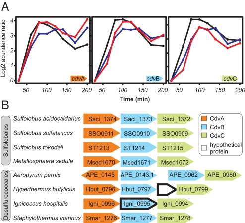
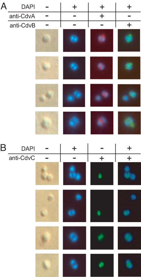
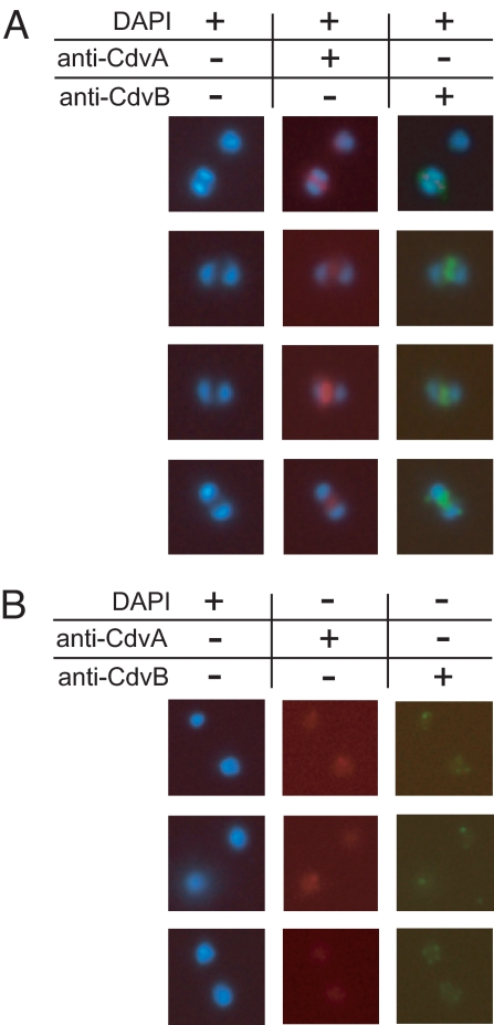
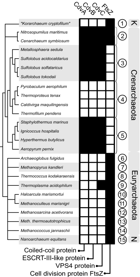
In contrast to the cell division machineries of bacteria, euryarchaea, and eukaryotes, no division components have been identified in the second main archaeal phylum, Crenarchaeota. Here, we demonstrate that a three-gene operon, cdv, in the crenarchaeon Sulfolobus acidocaldarius, forms part of a unique cell division machinery. The operon is induced at the onset of genome segregation and division, and the Cdv proteins then polymerize between segregating nucleoids and persist throughout cell division, forming a successively smaller structure during constriction. The cdv operon is dramatically down-regulated after UV irradiation, indicating division inhibition in response to DNA damage, reminiscent of eukaryotic checkpoint systems. The cdv genes exhibit a complementary phylogenetic range relative to FtsZ-based archaeal division systems such that, in most archaeal lineages, either one or the other system is present. Two of the Cdv proteins, CdvB and CdvC, display homology to components of the eukaryotic ESCRT-III sorting complex involved in budding of luminal vesicles and HIV-1 virion release, suggesting mechanistic similarities and a common evolutionary origin.


Conflict of interest statement

The authors declare no conflict of interest.

Figures

Fig. 1.
Fig. 1.
RNA abundance profiles and cdv operon structures. (A) Microarray-determined induction profiles over the cell cycle (6) for cdv genes in synchronized S. acidocaldarius cultures. Each graph represents an independent biological replicate. Initiation of genome segregation in the leading edge of the cell population was estimated to occur at ≈50–60 min, with cell division initiating 10–15 min later. (B) Conserved organization of cdv genes (arrows) across different crenarchaeal species. Only cdvA, cdvB, and cdvC orthologs that occur in operons are displayed. I. hospitalis gene product Igni_0995 displays distant homology to other CdvB proteins, indicated by dashed shading.
Fig. 2.
Fig. 2.
In situ immunofluorescence microscopy of S. acidocaldarius cells. Cultures were sampled in exponential growth phase. The first column depicts phase-contrast illumination of the cells shown in the consecutive columns. Nucleoids were stained with DAPI (4′,6-diamidino-2-phenylindole). Cdv proteins were stained with specific antibodies, followed by fluorescence visualization with Alexa Fluor-labeled secondary antibodies, as described in Materials and Methods. (A) Cells with two segregated nucleoids double-stained with anti-CdvA (red fluorescence) and anti-CdvB (green). (B) Anti-CdvC (green) stained cells. Note the absence of fluorescence signals in single-nucleoid cells (top two rows).
Fig. 3.
Fig. 3.
In situ immunofluorescence microscopy of S. acidocaldarius cells at different stages of genome segregation and constriction. Growth conditions and staining are specified in Materials and Methods, and in the legend to Fig. 2, respectively. (A) Cells that display a Cdv band together with a single nucleoid. (B) Cdv structures in cells undergoing constriction.
Fig. 4.
Fig. 4.
In situ immunofluorescence microscopy of exponentially growing S. acidocaldarius cells 6–8 h after antibiotic addition. Growth conditions, antibiotic concentrations, and staining are specified in the Material and Methods, and in the legend to Fig. 2, respectively. (A) Double-staining of tunicamycin-treated cells. (B) Double-staining of daunomycin-treated cells.
Fig. 5.
Fig. 5.
Phylogenetic distribution of division genes. Orthology-based representation of the phylogenetic distribution of cell division genes across sequenced archaeal genomes, based on the ArCOG database (31). Shaded and empty boxes represent the presence and absence of an ortholog in a given species, respectively. Abbreviations: 1, “Korarchaeota”; 2, Cenarchaeales; 3, Sulfolobales; 4, Thermoproteales; 5, Desulfurococcales; 6, Archaeoglobales; 7, Methanopyrales; 8, Thermococcales; 9, Thermoplasmatales; 10, Halobacteriales; 11, Methanomicrobiales; 12, Methanosarcinales; 13, Methanobacteriales; 14, Methanococcales; 15, Nanoarchaeales. N, Nanoarchaeota; K, Korarchaeota.

Comment in

References

    1. Woese CR, Kandler O, Wheelis ML. Towards a natural system of organisms: Proposal for the domains Archaea, Bacteria, and Eucarya. Proc Natl Acad Sci USA. 1990;87:4576–4579. - PMC - PubMed
    1. Lundgren M, Bernander R. Archaeal cell cycle progress. Curr Opin Microbiol. 2005;8:662–668. - PubMed
    1. Bernander R. The cell cycle of Sulfolobus. Mol Microbiol. 2007;66:557–562. - PubMed
    1. Margolin W. FtsZ and the division of prokaryotic cells and organelles. Nat Rev Mol Cell Biol. 2005;6:862–871. - PMC - PubMed
    1. Morgan DO. In: The Cell Cycle: Principles of Control. Lawrence E, editor. Sunderland: New Science Press Ltd.; 2007. pp. 157–173.

Publication types

MeSH terms