Comparative Study
. 2008 Nov 11;105(45):17420-5.
doi: 10.1073/pnas.0710725105. Epub 2008 Nov 5.

The parafibromin tumor suppressor protein inhibits cell proliferation by repression of the c-myc proto-oncogene

Affiliations
Comparative Study

The parafibromin tumor suppressor protein inhibits cell proliferation by repression of the c-myc proto-oncogene

Ling Lin et al. Proc Natl Acad Sci U S A. .

Abstract

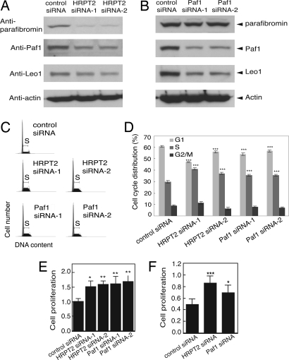
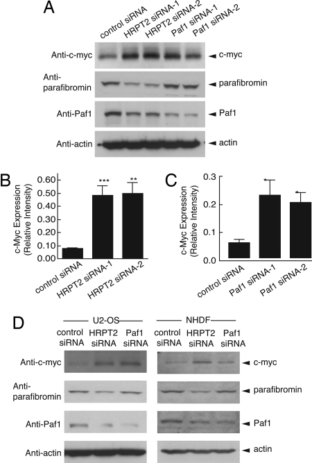
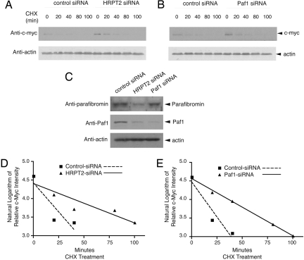
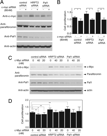
Parafibromin is a tumor suppressor protein encoded by HRPT2, a gene recently implicated in the hereditary hyperparathyroidism-jaw tumor syndrome, parathyroid cancer, and a subset of kindreds with familial isolated hyperparathyroidism. Human parafibromin binds to RNA polymerase II as part of a PAF1 transcriptional regulatory complex. The physiologic targets of parafibromin and the mechanism by which its loss of function can lead to neoplastic transformation are poorly understood. We show here that RNA interference with the expression of parafibromin or Paf1 stimulates cell proliferation and increases levels of the c-myc proto-oncogene product, a DNA-binding protein and established regulator of cell growth. This effect results from both c-myc protein stabilization and activation of the c-myc promoter, without alleviation of the c-myc transcriptional pause. Chromatin immunoprecipitation demonstrates the occupancy of the c-myc promoter by parafibromin and other PAF1 complex subunits in native cells. Knockdown of c-myc blocks the proliferative effect of RNA interference with parafibromin or Paf1 expression. These experiments provide a previously uncharacterized mechanism for the anti-proliferative action of the parafibromin tumor suppressor protein resulting from PAF1 complex-mediated inhibition of the c-myc proto-oncogene.


Conflict of interest statement

The authors declare no conflict of interest.

Figures

Fig. 1.
Fig. 1.
RNA interference with HRPT2 or Paf1 expression in human cells leads to down-regulation of endogenous Paf1 and Leo1 expression and increased cell proliferation. (A and B) Immunoblotting analysis of parafibromin (Top Panel), Paf1 (Second Panel), Leo1 (Third Panel) in HeLa cells following RNAi-mediated depletion of HRPT2 or Paf1 transcripts. Cells were treated with control siRNA or one of two HRPT2- or Paf1-specific siRNAs and analyzed as described in Methods. The expression of β-actin is shown as loading control (Fourth Panel). (C and D) Inhibition of parafibromin or Paf1 expression by RNAi resulted in increased S phase cell population. HeLa cells were treated with control siRNA or one of two HRPT2- or Paf1-specific siRNA and subjected to FACS analysis as described in Methods. (D) The histogram shows the distribution of different cell cycle population; values are representative of two independent experiments with similar results. Significance of the differences on S phase and G1 phase were evaluated by Student's t test (***, P < 0.001). (E and F) Proliferation analysis. Cultured HeLa (E) or NHDF cells (F) were treated as above for 48 h. Cell proliferation was estimated by a colorimetric cell proliferation assay. Presented are data representative of three individual repeats. Significance of the differences were evaluated by Student's t test (*** P < 0.005, **, P < 0.01, *, P < 0.05).
Fig. 2.
Fig. 2.
Up-regulation of c-myc protein in HeLa cells, U2-OS cells and NHDF cells following RNAi-mediated inhibition of HRPT2 or Paf1 expression. (A) HeLa cells were treated with control siRNA or one of two HRPT2- or Paf1-specific siRNA for 48 h and cells were then subjected to immunoblot analysis using anti-c-myc (Top Panel), anti-parafibromin (Second Panel), anti-Paf1 (Third Panel). (B and C) Based on Western blot analysis as presented in Fig. S7, quantification of c-myc expression was carried out by quantitative ECL and then normalized to actin expression. Significance of the differences were evaluated by Student's t test (***, P < 0.005, **, P < 0.01, *, P < 0.05). (D) U2-OS cells and NHDF cells were treated with control siRNA, HRPT2- or Paf1-specific siRNA as described in Methods. 48 h after treatment, cells were analyzed for expression of c-myc (Top Panel), parafibromin (Second Panel), Paf1 (Third Panel). In A and D the expression of β-actin is shown as a loading control.
Fig. 3.
Fig. 3.
Analysis of c-myc protein stability and half-life in response to inhibition of parafibromin or Paf1 expression. (A and B) Cultured HeLa cells were treated with control siRNA, HRPT2- or Paf1-specific siRNA. At 48 h after transfection, cycloheximide (CHX) was added to the culture medium and the cells were collected at 0, 20, 40, 80, and 100 min. Cell lysates were analyzed by immunoblotting using anti-c-myc or anti-actin antibodies. (C) The expression of parafibromin and/or Paf1 at the zero time point was analyzed by immunoblotting. The expression of β-actin is shown as a loading control. (D and E) Linear regression analysis of the natural logarithm of the relative intensity of c-myc protein expression in control and HRPT2- (D) or Paf1- (E) specific siRNA treated cells at different times of CHX treatment, quantified by quantitative ECL analysis and normalized to β–actin expression, is shown. In this analysis the y-value at time = 0 for all plots was 4.61 (the natural log of 100 [%] relative expression), and a y-value of 3.91 (indicating a relative expression of 50 [%]) maps to the time corresponding to the c-myc protein half-life.
Fig. 4.
Fig. 4.
c-Myc is downstream of the Paf1 complex in the control of cell proliferation. (A) c-myc expression in HeLa cells treated with 80 nM of c-myc siRNA in the presence of control, HRPT2- or Paf1-specific siRNA. Efficacy of RNAi-mediated gene silencing of c-myc (Top Panel), HRPT2 (Second Panel) and Paf1 (Third Panel) were assessed by immunoblot analysis. (B) For the cell proliferation assay, HeLa cells were treated with control siRNA, HRPT2- or Paf1-specific siRNA in the absence or presence of 80 nM of c-myc-specific siRNA. Forty-eight hours after treatment, proliferation was estimated by a colorimetric assay as described in Methods. Data are representative of three individual repeats with similar results. (C) c-Myc expression in HeLa cells treated with control-, HRPT2- or Paf1-specific siRNA in the presence of 0, 20, or 40 nM of c-myc-specific siRNA. Gene silencing efficiency of c-myc (Top Panel), HRPT2 (Second Panel) and Paf1 (Third Panel) are monitored by Western blot analysis. (D) Cell proliferation of HeLa cells after treatment for 48h with HRPT2- or Paf1-specific siRNA in the presence of 0, 20, or 40 nM of c-myc-specific siRNA was estimated by a colorimetric assay. Data are representative of three independent experiments with similar results. Significance of the differences were evaluated by Student's t test (**, P < 0.01, *, P < 0.05). In A and C the expression of β-actin is shown as a loading control.

References

    1. Carpten JD, et al. HRPT2, encoding parafibromin, is mutated in hyperparathyroidism-jaw tumor syndrome. Nat Genet. 2002;32:676–680. - PubMed
    1. Jackson CE, et al. Hereditary hyperparathyroidism and multiple ossifying jaw fibromas: A clinically and genetically distinct syndrome. Surgery. 1990;108:1006–1012. - PubMed
    1. Chen JD, et al. Hyperparathyroidism-jaw tumour syndrome. J Intern Med. 2003;253:634–642. - PubMed
    1. Simonds WF, et al. Familial isolated hyperparathyroidism is rarely caused by germline mutation in HRPT2, the gene for the hyperparathyroidism-jaw tumor syndrome. J Clin Endocrinol Metab. 2004;89:96–102. - PubMed
    1. Shattuck TM, et al. Somatic and germ-line mutations of the HRPT2 gene in sporadic parathyroid carcinoma. N Engl J Med. 2003;349:1722–1729. - PubMed

Publication types

LinkOut - more resources