. 2008 Nov 11;105(45):17379-83.
doi: 10.1073/pnas.0807764105. Epub 2008 Nov 5.

Structure of transmembrane pore induced by Bax-derived peptide: evidence for lipidic pores

Affiliations

Structure of transmembrane pore induced by Bax-derived peptide: evidence for lipidic pores

Shuo Qian et al. Proc Natl Acad Sci U S A. .

Abstract

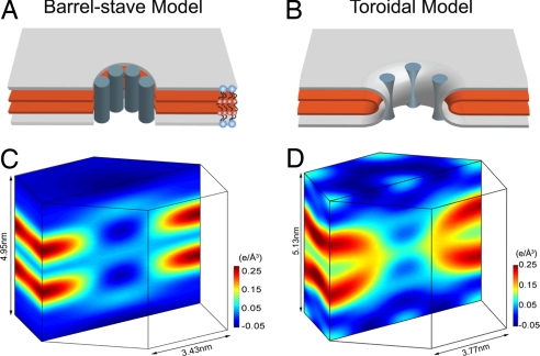
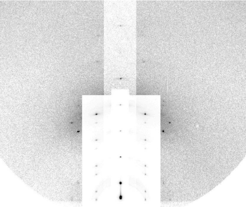
The structures of transmembrane pores formed by a large family of pore-forming proteins and peptides are unknown. These proteins, whose secondary structures are predominantly alpha-helical segments, and many peptides form pores in membranes without a crystallizable protein assembly, contrary to the family of beta-pore-forming proteins, which form crystallizable beta-barrel pores. Nevertheless, a protein-induced pore in membranes is commonly assumed to be a protein channel. Here, we show a type of peptide-induced pore that is not framed by a peptide structure. Peptide-induced pores in multiple bilayers were long-range correlated into a periodically ordered lattice and analyzed by X-ray diffraction. We found the pores induced by Bax-derived helical peptides were at least partially framed by a lipid monolayer. Evidence suggests that the formation of such lipidic pores is a major mechanism for alpha-pore-forming proteins, including apoptosis-regulator Bax.


Conflict of interest statement

The authors declare no conflict of interest.

Figures

Fig. 1.
Fig. 1.
Two models of peptide-induced pores in membranes and the experimental proof for each. (A and B) Schematics of the barrel-stave model (A) and the toroidal model (B) are shown in a 3D view cut through the pores, showing only the layers representing the lipid headgroups (silver) and the Br atoms (red). Blue-gray cylinders represent peptide segments; the actual distribution and orientation of peptides are expected to vary with peptide and lipid. Schematic lipid molecules [1,2-distearoyl(9–10dibromo)-sn-glycero-3-phosphocholine] are shown in A, where the red dots represent the Br atoms on the chains. (C and D) The normalized electron density distributions of Br atoms in a unit cell of the R phase containing alamethicin and Bax-α5, respectively. (The data for C were reproduced from ref. .) Br atoms are distributed in the high-density (yellow-red-black) region. The nonuniformity in the low-density region (light blue to dark blue, which average to ≈0 e/Å3) is due to the limited resolution (28).
Fig. 2.
Fig. 2.
Grazing-angle diffraction pattern from the R phase of a Bax-α5 and di18:0(9,10Br)PC mixture in the molar ratio of 1:30 at 60% RH, 25 °C. The vertical (qz) is normal to the substrate. To show all peaks clearly, intensity attenuators were applied to the peaks on qz and on the 2 columns next to qz.
Fig. 3.
Fig. 3.
Multiwavelength anomalous diffraction analyses for the detected peaks. For each independent peak, the square root of the integrated intensity, ∣Fλ∣, is plotted as a function of ∣f′λ∣/fn. The data are fit with a straight line, from which ∣F0∣, ∣F2∣, and the ratio ∣F0∣/∣F2∣ are obtained. The results are in Table 1.
Fig. 4.
Fig. 4.
Three-dimensional swelling method for phase determination applied to the rhombohedral phase (42). Shown is the swelling method applied to different qr series using the diffraction amplitudes obtained at 50% RH (open circles) and 60% RH (filled circles). (A) qr = 0 nm−1 series. (B) qr = 1.11 nm−1 series. (C) qr = 1.92 nm−1 series. The vertical scales are relative. The solid curves are F(qH,K,qz) of equation 12 in ref. constructed from 1 set of data measured at 1 relative humidity (see details in ref. 42).

References

    1. Panchal RG, Smart ML, Bowser DN, Williams DA, Petrou S. Pore-forming proteins and their application in biotechnology. Curr Pharm Biotechnol. 2002;3:99–115. - PubMed
    1. Heuck AP, Tweten R K, Johnson AE. Beta-barrel pore-forming toxins: Intriguing dimorphic proteins. Biochemistry. 2001;40:9065–73. - PubMed
    1. Menestrina G, Serra MD, Lazarovici P. Pore-forming Peptides and Protein Toxins. London: Taylor & Francis; 2003. pp. 1–315.
    1. van der Goot FG. Current Topics in Microbiology and Immunology. Vol 257. Berlin: Springer; 2001. Pore-forming toxins; pp. 1–166.
    1. Zasloff M. Antimicrobial peptides of multicellular organisms. Nature. 2002;415:389–95. - PubMed

Publication types

MeSH terms