. 2009 Jan;20(1):256-69.
doi: 10.1091/mbc.e08-03-0288. Epub 2008 Nov 5.

Alternative requirements for Vestigial, Scalloped, and Dmef2 during muscle differentiation in Drosophila melanogaster

Affiliations

Alternative requirements for Vestigial, Scalloped, and Dmef2 during muscle differentiation in Drosophila melanogaster

Hua Deng et al. Mol Biol Cell. 2009 Jan.

Abstract

Vertebrate development requires the activity of the myocyte enhancer factor 2 (mef2) gene family for muscle cell specification and subsequent differentiation. Additionally, several muscle-specific functions of MEF2 family proteins require binding additional cofactors including members of the Transcription Enhancing Factor-1 (TEF-1) and Vestigial-like protein families. In Drosophila there is a single mef2 (Dmef2) gene as well single homologues of TEF-1 and vestigial-like, scalloped (sd), and vestigial (vg), respectively. To clarify the role(s) of these factors, we examined the requirements for Vg and Sd during Drosophila muscle specification. We found that both are required for muscle differentiation as loss of sd or vg leads to a reproducible loss of a subset of either cardiac or somatic muscle cells in developing embryos. This muscle requirement for Sd or Vg is cell specific, as ubiquitous overexpression of either or both of these proteins in muscle cells has a deleterious effect on muscle differentiation. Finally, using both in vitro and in vivo binding assays, we determined that Sd, Vg, and Dmef2 can interact directly. Thus, the muscle-specific phenotypes we have associated with Vg or Sd may be a consequence of alternative binding of Vg and/or Sd to Dmef2 forming alternative protein complexes that modify Dmef2 activity.


Figures

Figure 1.
Figure 1.
sd in situ reveals the expression of sd in both SMs and dorsal vessel. (A1–A3) The specificity of the sd probe for in situ was tested on wing disk, and sd expression pattern in wing disk was accurately revealed by this probe. (B) sd transcript was found mostly in the heart region of the dorsal vessel (dashed line). It also appears in the hind gut (arrow). (C1–C3) sd transcript was detected in SMs of stage 13 wild-type embryos. SMs are visualized by Dmef2 staining. sd transcript was also detected in the CNS cells (arrows). (D1–D3) It failed to detect sd transcript in SMs of wild-type embryos at early stage 16. High expression of sd was found in salivary gland at this stage (arrow).
Figure 2.
Figure 2.
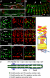
sd, vg, and Dmef2 are coexpressed in embryonic muscles. To facilitate double staining, sd expression was detected by examining sd reporter constructs (sdETX4, A and A′), or using 3xFLAG-Sd driven by sd-GAL4 (B–E). Muscle cells are marked with anti-Dmef2 (green in A and B), and Vg is labeled by anti-Vg (green in C–E). 3xFLAG-Sd and LacZ are visualized with anti-FLAG and anti-β-Gal (red), respectively. (A) In stage 16 embryos the sdETX4 reporter is activated in the heart region of the dorsal vessel and in some cardiac cells in the aorta region (arrowheads). It is also expressed in the hind gut, underneath the visceral muscles (VMs, small arrowhead). (A) A dorsal-lateral view; (A′) a dorsal view. (B) sd-GAL4 drives expression of 3xFLAG-Sd in several cardiac cells (arrows) and ∼31% cells of somatic muscles (SMs, arrowheads) at stage 13. Note that sd-GAL4 is also activated in cells of central neuron system (CNS, empty arrow). Dmef2 is present in all muscle cells. C1–C3 shows the dorsal SMs where vg is expressed at stage 13. 3xFLAG-Sd can be detected in some SMs. D1–D3 shows that vg is expressed in the DA3, LL1, and VL1-4 muscles when 3xFLAG-Sd appears in all SMs at stage 16. Vg also appears in some neuronal cells (arrowheads). DA1-2 are not shown because they are out of the field of view. E1–E3 shows that vg is still expressed in the DA3, LL1, and VL1-4 muscles when the expression of 3xFLAG-Sd fades in SMs and appears only in some ventral SMs at late stage 16. At this stage, 3xFLAG-Sd appears with Vg in the neuron cells shown above (arrowheads). (F) A schematic drawing of a stage 16 embryonic dorsal vessel (dorsal view, anterior to the left). Heart cells include two parallel rows of Dmef2-positive cardiac cells in the middle with four Tinman-positive cardiac cells per hemisegment starting from T1. Dmef2-negative pericardial cells surround the cardiac cells. On the right is a schematic representation of the embryonic SMs in each abdominal hemisegment A2–A7 (lateral view with anterior up) using the nomenclature of Crossley (1978). Inner, middle, and outer muscle layers are shown in yellow, blue, and red, respectively (Bate and Rushton, 1993).
Figure 3.
Figure 3.
The sd3L and vgnull mutants have defects in embryonic muscle development. Muscle cells are marked with anti-Dmef2 (green), and muscle fibers are visualized by phalloidin staining (red). Anterior is to the left. (A) In stage 13 wild-type embryos (A′ is the close-up of the boxed area in A), there are six cardiac cells per hemisegment (A′). (B) In stage 13 sd3Lmutant embryos (B′ is the close-up of boxed area in B), there are many cardiac cells with enlarged nuclei (arrowheads) relative to neighboring cells (arrows), and there are fewer cardiac cells per hemisegment (compare A′ with B′). (C) The number of cardiac cells on one side of sd3L embryos (41 ± 4.7, mean ± Sd, n = 8) is less than that of wild type (52 ± 0, n = 10). (D) The SMs of a wild-type embryo at early stage 16. (E) The SMs of a sd3Lmutant embryo at the same stage. Many ventral SMs (VO4-6, see Figure 2) have severe developmental defects or are absent entirely (arrows, compare E with D). (F) Actin was stained by phalloidin in a stage 16 wild-type embryo. The VO4-6 muscles are indicated by a bracket and the VL2 muscle is demarked by an asterisk. (G) In the sd3L mutant, the VO4-6 muscles is absent in some segments (bracket). (H) In the vgnull mutant, the VL2 muscle is absent in some segments (star), and the VO1 muscle underneath can be seen (arrow).
Figure 4.
Figure 4.
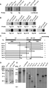
Interactions between Sd, Vg, and Dmef2 can be shown by coimmunoprecipitation (CoIP) assays. Indicated proteins with different tags were coexpressed in S2 cells, and CoIP was performed using anti-FLAG beads in both control and experiment samples. Proteins coming down with the beads and the relative expression level of the proteins in the lysate were detected with corresponding tag antibodies. (A) Dmef2 and Vg were coIPed simultaneously with Sd (arrows). In the control, tagged Vg and Demf2 did not come down with the beads (arrowheads). There are additional bands for Vg and Dmef2, likely because of posttranslational modifications. (B) Sd coIPed Vg or Dmef2 without coexpression of Dmef2 or Vg, respectively. (C) The anti-FLAG bead-purified IP complex of 3xFLAG-Sd and 6xMyc-Dmef2 and that of 3xFLAG-Dmef2 and 6xMyc-Sd were immunoblotted with anti-Myc and anti-Vg antibodies. Significant levels of Vg could not be detected in these complexes. Arrows show the proteins coming down with the beads, and arrowheads show the primary antibody bands. (D) CoIP was performed on S2 cells transfected with the indicated proteins at different times, following heat shock. The relative amount of coIPed Dmef2 (right) increased with the expression level of Vg and Dmef2 (left).
Figure 5.
Figure 5.
Vg can interact with Dmef2 at a different site than it interacts with Sd. (A) A positive control shows an interaction with a known Vg binding partner (GST-Sd). A similar robust interaction is detected between Vg and GST-Dmef2. Luciferase serves as a negative control. (B) Two separate domains (illustrated with boxes in C) in Vg are capable of interacting with Dmef2. All deletions except Vg3-9 interact with GST-Dmef2 because at least one of the two domains is intact in all other deletions tested. (D and E) Gel analysis confirming expression of proteins used in pulldown assays (arrowheads). Protein size is indicated on the left. SID, Sd interaction domain; TAD, transcription activation domain.
Figure 6.
Figure 6.
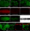
The Sd–Vg complex represses Dmef2 function during muscle differentiation. Embryos (stage 16) are shown as lateral views, and dorsal is up, with anterior to the left. (A) The three constrictions that subdivide the midgut into four chambers are shown with arrows in a wild-type embryo. (B) Ectopic expression of Vg in visceral muscles of embryos via Dmef2-GAL4 does not affect the formation of these constrictions. (C) Ectopic coexpression of Sd and Vg in visceral muscles leads to the repression of Dmef2 function in these muscles and all three constrictions disappear. (D–F) The Myosin staining of wild-type embryos (D) and the embryos overexpressing Sd or Sd and Vg (E and F). The apparent level of myosin staining is reduced when Sd is overexpressed in Dmef2-expressing muscles (E) and even more reduced when Vg and Sd are present (F). (G and H) Actin staining by phalloidin failed to show the formation of myofibers in muscles of embryo overexpressing Sd and Vg. (I) The results of RT-PCR from stage 12–15 wild-type embryos or embryos overexpressing Sd and Vg. The relative amount of act57B and mhc mRNA in embryos overexpressing Sd and Vg is much lower than wild type. rp49 mRNA was used as loading control. (J) Ventral SMs in one segment are visualized by actin staining in a wild-type embryo, and VL1 is shown by the dashed frame. (K1–K3) Specific overexpression of Sd and Vg in VL1 via 5053-GAL4 leads to the missing of myofiber in this muscle. (Sd and Vg are 3xFLAG tagged and 3xHA tagged, respectively).
Figure 7.
Figure 7.
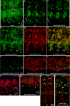
Ectopic-expression of Sd and/or Vg via Dmef2-GAL4 leads to abnormal development of somatic muscles. Embryos (stage 16) are shown as lateral views, and dorsal is up, with anterior to the left. (A) Muscle fibers are visualized by anti-β-Gal (green) in control embryos expressing LacZ in cytoplasm under the control of the act57B promoter shown in A. Arrows point to muscle LL1, VL1, and VL2, and the bracket shows the ventral oblique muscles (VO4-6, see Figure 3D). VO4-6 muscles produce three projections that expand posterior-ventrally. (B) Muscle cells are marked with anti-Dmef2 (green) in wild-type embryos. Bracket shows muscle VO4-6. (C) In embryos ectopically expressing 3xHA-Vg, the extension of VO4-6 is lost (arrows), but the DMs are still highly organized (compare B with C). (D1–D3) Embryos (act57B-lacZ) ectopically expressing 3xFLAG-Sd (red) have disorganized somatic muscles. LacZ staining shows the whole muscle and FLAG staining shows the muscle nuclei. Muscle LL1, VL1, and VL2 do not develop well or disappear in some segments (arrows). The ventral muscle VO4-6 can still produce projections that expand ventrally, but there are more projections than wild type, and some projections expand anterior-ventrally (arrowheads, compare D1 with A). (E1–E3) Embryos (act57B-lacZ) ectopically expressing both 3xFLAG-Sd (red) and 3xHA-Vg have disorganized SMs, and the extension of VO4-6 is also lost (arrows). The expression level of LacZ is generally very low compared with the control, and cells with high expression levels usually do not express 3xFLAG-Sd or have low expression (arrowheads). Staining of 3xHA-Vg is not shown, because 3xFLAG-Sd and 3xHA-Vg always appear in the same muscle cells. (F) Wild-type ventral SMs (arrows) are labeled by phalloidin, and the segment border is labeled with anti-βPS-integrin. Arrows point to muscle VO4-6 and VA3. (G) In embryos overexpressing 3xHA-Vg, VL1-4 muscles are not affected, but ventral SMs are severely affected. It seems that these muscles are still there, but their migrations are either inhibited (arrowhead) or directed in a different path (arrows), which lead to no long extensions of muscle fiber. HA staining (green) shows the nuclei of muscles. (H) Wild-type SMs are labeled with anti-Dmef2 (red), and the segment border is labeled with anti-βPS-integrin. All muscle cells have their proper positions relative to the border (arrowheads). (I) In embryos overexpressing both 3xFLAG-Sd and 3xHA-Vg, muscle cells lose their positions and appear to cluster along the segments border (arrowheads). Large gaps are seen within each segment (arrows).
Figure 8.
Figure 8.
Functional interactions between Sd, Vg, and Dmef2 can be shown by ectopic expression of various combinations of Sd, Vg, and Dmef2 in heart cells. Embryos shown as lateral views and dorsal up with anterior to the left are at stage 14 and stained with antibodies as indicated by the colored lettering. Brackets show the area where the heart cells are located. (A′) Dmef2-GAL4 drives the expression of LacZ in all SMs and in both cardiac cells and pericardial cells (arrows). (A and B) Wild type. There is one row of Dmef2-positive cardiac cells (A) and four Tin-positive cardiac cells per hemisegment (B, arrows). (C and D) Embryos ectopic-expressing 3xHA-Vg have the normal one row of Dmef2-positive cardiac cells (C), but now have six Tin-positive cardiac cells per hemisegment (D, arrows). (E and F) Embryos ectopic-expressing 3xFLAG-Sd have two to three rows of Dmef2-positive cardiac cells (E). Tin-positive heart cells become disorganized: sometimes, you see only two Tin-positive cardiac cells in one hemisegment (F, arrows) and sometimes, you see Tin-positive cells appear in the region of the SMs (F, arrowhead). (G and H) Embryos overexpressing 6xMyc-Dmef2 have two rows of Dmef2-positive cardiac cells (G), but there are four Tin-positive cardiac cells per hemisegment like wild type (H, arrows). (I and J) Embryos overexpressing both 6xMyc-Dmef2 and 3xHA-Vg have two to three rows of Dmef2-positive cardiac cells (I) and six Tin-positive cardiac cells per hemisegment (J, arrows). Many more Tin-positive heart cells also appear (compared J with D and H). (K–M) Embryos overexpressing both 3xFLAG-Sd and 6xMyc-Dmef2. The phenotype is similar to E and F, but heart cells are more organized (compare M with F). Arrowheads show the Tin-positive cells mixed with SMs. (N and O) Embryos overexpressing both 3xFLAG-Sd and 3xHA-Vg have only a few Dmef2-positive cardiac cells left (N, arrows) and all the Tin-positive cells appear in the region of SMs (O, arrowheads).

References

    1. Azpiazu N., Frasch M. tinman and bagpipe: two homeo box genes that determine cell fates in the dorsal mesoderm of Drosophila. Genes Dev. 1993;7:1325–1340. - PubMed
    1. Bao M. Z., Schwartz M. A., Cantin G. T., Yates J. R., 3rd, Madhani H. D. Pheromone-dependent destruction of the Tec1 transcription factor is required for MAP kinase signaling specificity in yeast. Cell. 2004;119:991–1000. - PubMed
    1. Bate M., Rushton E. Myogenesis and muscle patterning in Drosophila. C. R. Acad. Sci. III. 1993;316:1047–1061. - PubMed
    1. Baylies M. K., Bate M., Ruiz Gomez M. Myogenesis: a view from Drosophila. Cell. 1998;93:921–927. - PubMed
    1. Bernard F., Lalouette A., Gullaud M., Jeantet A. Y., Cossard R., Zider A., Ferveur J. F., Silber J. Control of apterous by vestigial drives indirect flight muscle development in Drosophila. Dev. Biol. 2003;260:391–403. - PubMed

Publication types

MeSH terms