. 2009 Jan;20(1):389-99.
doi: 10.1091/mbc.e07-11-1132. Epub 2008 Nov 5.

Molecular clustering of STIM1 with Orai1/CRACM1 at the plasma membrane depends dynamically on depletion of Ca2+ stores and on electrostatic interactions

Affiliations

Molecular clustering of STIM1 with Orai1/CRACM1 at the plasma membrane depends dynamically on depletion of Ca2+ stores and on electrostatic interactions

Nathaniel Calloway et al. Mol Biol Cell. 2009 Jan.

Abstract

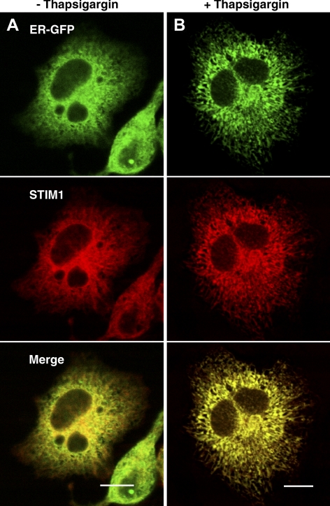
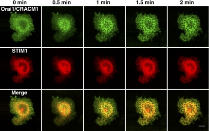
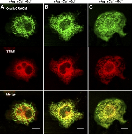
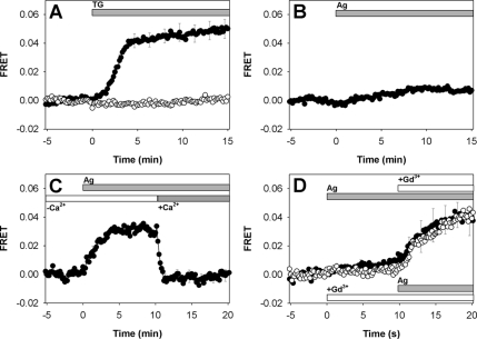
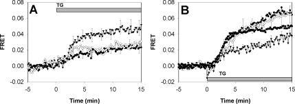
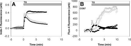
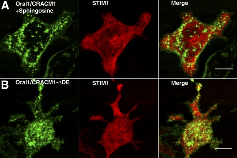
Activation of store operated Ca(2+) entry involves stromal interaction molecule 1 (STIM1), localized to the endoplasmic reticulum (ER), and calcium channel subunit (Orai1/CRACM1), localized to the plasma membrane. Confocal microscopy shows that thapsigargin-mediated depletion of ER Ca(2+) stores in RBL mast cells causes a redistribution of STIM1, labeled with monomeric red fluorescent protein (mRFP), to micrometer-scale ER-plasma membrane junctions that contain Orai1/CRACM1, labeled with monomeric Aequorea coerulescens green fluorescent protein (AcGFP). Using fluorescence resonance energy transfer (FRET), we determine that this visualized coredistribution is accompanied by nanoscale interaction of STIM1-mRFP and AcGFP-Orai1/CRACM1. We find that antigen stimulation of immunoglobulin E receptors causes much less Orai1/CRACM1 and STIM1 association, but strong interaction is observed under conditions that prevent refilling of ER stores. Stimulated association monitored by FRET is inhibited by sphingosine derivatives in parallel with inhibition of Ca(2+) influx. Similar structural and functional effects are caused by mutation of acidic residues in the cytoplasmic segment of Orai1/CRACM1, suggesting a role for electrostatic interactions via these residues in the coupling of Orai1/CRACM1 to STIM1. Our results reveal dynamic molecular interactions between STIM1 and Orai1/CRACM1 that depend quantitatively on electrostatic interactions and on the extent of store depletion.


Figures

Figure 1.
Figure 1.
RBL mast cell expressing STIM1-mRFP (red) and ER-eGFP (green) exhibits stimulation-dependent changes in localization of these proteins. Representative cells are shown before (A) and after (B) stimulation with thapsigargin. Note the extension of the ER network throughout the cytoplasm after stimulation and the increased colocalization of STIM1 with the luminal ER marker. Bars, 10 μm.
Figure 2.
Figure 2.
Time course of AcGFP-Orai1/CRACM1 (green) and STIM1-mRFP (red) formation of colabeled plasma membrane domains after thapsigargin stimulation. The patches seen in the last image, formed after 2 min of stimulation, remained relatively unchanged for an additional 10 min (not shown). Dorsal cell surface images. Bar, 10 μm.
Figure 3.
Figure 3.
Confocal images of AcGFP-Orai1/CRACM1 (green) and STIM1-mRFP (red) in dorsal slices of RBL mast cells stimulated with antigen under different conditions affecting calcium influx. Cell stimulated in BSS buffer with (A) and without (B) CaCl2. (C) Cell stimulated in BSS with Ca2+ and 6 μM GdCl3. Bars, 10 μm.
Figure 4.
Figure 4.
FRET between AcGFP-Orai1/CRACM1 and STIM1-mRFP in plasma membrane of RBL mast cells. (A) FRET after addition of 150 nM thapsigargin (indicated by bar) to labeled cells (solid circles; error bars show SEM) compared with control cells expressing PH-eGFP and unlabeled Orai1/CRACM1 instead of AcGFP-Orai1/CRACM1 (open circles, SEM ∼2.0 × 10⁻³). (B) FRET after antigen stimulation with 3 nM DNP-BSA in BSS (SEM ∼1.0 × 10⁻³). (C) FRET after same antigen stimulation but in the absence of extracellular Ca2+. CaCl2 (1.8 mM) was restored as indicated by bar (error bars show SEM). (D) FRET after stimulation by antigen followed by addition of 6 μM GdCl3 (solid circles; top bars; error bars show SEM; solid line) or after GdCl3 addition followed by antigen stimulation (open circles; bottom bars; error bars omitted; dotted line).
Figure 5.
Figure 5.
(A) Thapsigargin-stimulated FRET between AcGFP-Orai1/CRACM1 and STIM1-mRFP in the presence of d-sphingosine (7.6 μM, open circles), DMS (7.6 μM, closed circles; error bars omitted), and TMS (7.6 μM, triangles). (B) Thapsigargin-stimulated FRET between STIM1-mRFP and AcGFP-Orai1/CRACM1 (squares, same as Figure 4A, shown for comparison; error bars omitted), AcGFP-Orai1/CRACM1ΔD (closed circles; error bars omitted), AcGFP-Orai1/CRACM1ΔE (open circles), and AcGFP-Orai1/CRACM1ΔDE (triangles). Error bars show SEM.
Figure 6.
Figure 6.
(A) Effects of sphingosine derivatives on thapsigargin induced Ca2+ mobilization, indo-1-monitored. Ca2+ response of suspended RBL cells for untreated cells (closed circles), cells in the presence of d-sphingosine (7.6 μM, open circles), DMS (7.6 μM, closed triangles), TMS (7.6 μM, open triangles). Every 10th data point is shown for clarity. (B) Calcium measurements of COS7 cells monitored by fluo-4 imaging. Thapsigargin mediated Ca2+ mobilization is shown for untransfected cells (closed circles), cells expressing STIM1-mRFP with AcGFP-Orai1/CRACM1 (open circles), AcGFP-Orai1/CRACM1ΔE (open triangles), and AcGFP-Orai1/CRACM1ΔDE (closed triangles).
Figure 7.
Figure 7.
(A) AcGFP-Orai1/CRACM1 in the presence of 7.6 μM d-sphingosine exhibits plasma membrane patches without STIM1-mRFP redistribution. (B) AcGFP-Orai1/CRACM1ΔDE and STIM1-mRFP exhibits plasma membrane patches without STIM1-mRFP redistribution. Compare these dorsal surface images to Figure 3, t = 0. Bars, 10 μm.

References

    1. Baba Y., et al. Coupling of STIM1 to store-operated Ca2+ entry through its constitutive and inducible movement in the endoplasmic reticulum. Proc. Natl. Acad. Sci. USA. 2006;103:16704–16709. - PMC - PubMed
    1. Baba Y., Nishida K., Fujii Y., Hirano T., Hikida M., Kurosaki T. Essential function for the calcium sensor STIM1 in mast cell activation and anaphylactic responses. Nat. Immunol. 2008;9:81–88. - PubMed
    1. Barr V. A., et al. Dynamic movement of the calcium sensor STIM1 and the calcium channel orai1 in activated T-cells: puncta and distal caps. Mol. Biol. Cell. 2008;19:2802–2817. - PMC - PubMed
    1. Campbell R. E., Tour O., Palmer A. E., Steinbach P. A., Baird G. S., Zacharias G. S., Tsien R. Y. A monomeric red fluorescent protein. Proc. Natl. Acad. Sci. USA. 2002;99:7877–7882. - PMC - PubMed
    1. Das R., Hammond S., Holowka D., Baird B. Real-time cross-correlation image analysis of early events in IgE receptor signaling. Biophys. J. 2008;94:4996–5008. - PMC - PubMed

Publication types

MeSH terms

LinkOut - more resources