. 2009 Apr 2;113(14):3245-53.
doi: 10.1182/blood-2008-07-166389. Epub 2008 Nov 5.

The transcription factor c-Myc enhances KIR gene transcription through direct binding to an upstream distal promoter element

Affiliations

The transcription factor c-Myc enhances KIR gene transcription through direct binding to an upstream distal promoter element

Frank Cichocki et al. Blood. .

Abstract

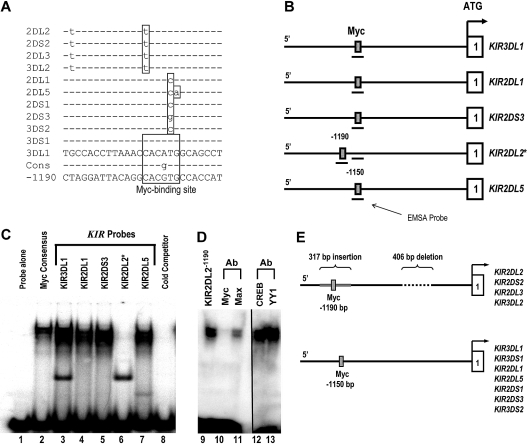
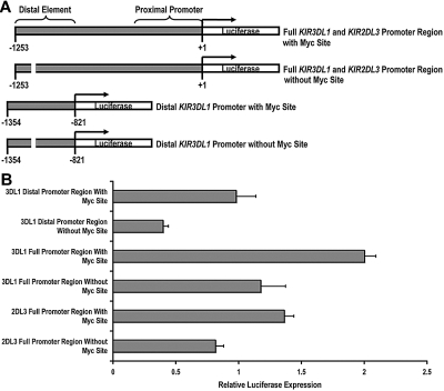
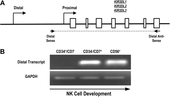
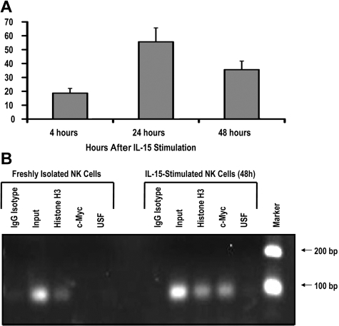
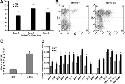
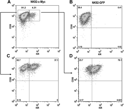
The killer cell immunoglobulin-like receptor (KIR) repertoire of natural killer (NK) cells determines their ability to detect infected or transformed target cells. Although epigenetic mechanisms play a role in KIR gene expression, work in the mouse suggests that other regulatory elements may be involved at specific stages of NK-cell development. Here we report the effects of the transcription factor c-Myc on KIR expression. c-Myc directly binds to, and promotes transcription from, a distal element identified upstream of most KIR genes. Binding of endogenous c-Myc to the distal promoter element is significantly enhanced upon interleukin-15 (IL-15) stimulation in peripheral blood NK cells and correlates with an increase in KIR transcription. In addition, the overexpression of c-Myc during NK-cell development promotes transcription from the distal promoter element and contributes to the overall transcription of multiple KIR genes. Our data demonstrate the significance of the 5' promoter element upstream of the conventional KIR promoter region and support a model whereby IL-15 stimulates c-Myc binding at the distal KIR promoter during NK-cell development to promote KIR transcription. This finding provides a direct link between NK-cell activation signals and KIR expression required for acquisition of effector function during NK-cell education.


Figures

Figure 1
Figure 1
c-Myc binds to the distal KIR promoter element in NK cells. (A) The intergenic region preceding each of the KIR genes on chromosome 19 was analyzed for transcription factor binding sites. The sequence of the distal promoter element, −1150 bp upstream of the translational start site, was aligned for each KIR gene, and a predicted Myc-binding site was identified. The sequence of the putative Myc-binding region of the KIR3DL1 gene is shown, and polymorphisms found in the other genes are highlighted, as well as the consensus Myc probe used for EMSA analysis (Cons). The sequence of the distal promoter element −1190 bp upstream of the translational start site for the KIR2DL2/2DS2/2DL3/3DL2 is also shown. (B) The location of the EMSA probes spanning the Myc-binding sequence 1150 bp upstream of selected KIR translational start sites. The location of the EMSA probe spanning the consensus Myc site 1190 bp upstream of the KIR2DL2 translational start site is also shown. (C) EMSA analysis of the predicted Myc-binding sites. Probes corresponding to the nucleotide sequences shown in panel A were used for an EMSA with YT cell extracts. A Myc consensus probe alone control is shown in lane 1. Complexes formed by individual 32P-labeled KIR probes are shown in lanes 2 to 7. A cold competitor control for probe specificity is shown in lane 8. (D) Inhibition of the complex formed by the KIR2DL21190 probe with anti-c-Myc and anti-Max antibodies is shown in lanes 10 and 11. The vertical line has been inserted between lanes 11 and 12 to indicate a repositioned gel lane. Inhibition of the complex formed by the KIR2DL21190 probe with anti-CREB and anti-YY1 control antibodies is shown in lanes 12 and 13. (E) Because of a 406-bp deletion and a 317-bp Alu insertion in the intergenic region of KIR2DL2/2DS2/2DL3/3DL2, a Myc-binding consensus is located at position −1190 relative to the transcriptional start site instead of −1150, where the site is located for the rest of the KIR genes.
Figure 2
Figure 2
c-Myc acts in a direct and specific manner at the distal promoter element to influence KIR transcription. (A) Schematics of the full-length and distal promoter fragments with and without the Myc site that were cloned into the pGL3 basic reporter vector. (B) Each pGL3 vector containing full and distal promoter sequences was electroporated into the NKL-cell line, and luciferase expression was determined 6 hours after transfection. Results represent the mean luciferase levels normalized to Renilla signals from 3 independent experiments.
Figure 3
Figure 3
Full-length KIR transcripts originate from the distal promoter region during NK-cell development. (A) PCR primers were designed to amplify full-length KIR2DL1, -2DL2, and -2DL3 transcripts originating from the distal promoter region. The amplified region begins upstream of the classical transcriptional start site (distal sense) and extends to the stop codon (distal antisense). Open boxes represent KIR gene exons. (B) Mononuclear cells from umbilical cord blood were sorted for specific NK-cell precursor populations based on the expression of the developmental markers CD34, CD7, and CD56. Sorted cells were analyzed for the presence of distal transcripts by RT-PCR using the primers shown in panel A. Control PCRs were carried out using GAPDH primers to confirm the presence of cDNA.
Figure 4
Figure 4
IL-15 increases c-Myc transcription and c-Myc binding to the KIR distal promoter element. (A) KIR-negative NK cells were isolated from adult peripheral blood and cultured in the presence of 10 ng/mL IL-15 for 4, 24, or 48 hours. Cells were harvested at each time point, and c-myc transcript levels were determined by quantitative RT-PCR (n = 3). Samples are normalized to unstimulated peripheral blood NK-cell controls. (B) ChIP analysis of c-Myc binding to freshly isolated KIR-negative NK cells or NK cells stimulated with 10 ng/mL IL-15 for 48 hours. Input sample represents the total input DNA contained in the chromatin aliquot used for each ChIP. Rabbit antibodies used for the IP were purified rabbit IgG (as a negative control), histone H3 (as a positive control), anti-Myc, and anti-USF. Results from a 35-cycle PCR are shown.
Figure 5
Figure 5
IL-15 induces KIR expression in KIR-negative NK cells. KIR-negative NK cells were isolated from adult peripheral blood and stimulated with 10 ng/mL IL-15 for 48 to 120 hours. (A) FACS analysis was performed at baseline before culture and 120 hours after IL-15 stimulation staining with a cocktail of APC-conjugated NCAM16.2 and PE-conjugated DX9, EB6, GL183, and FES172 monoclonal antibodies (n = 9). (B) Proximal coding KIR transcript and distal KIR transcript levels were measured after 48 and 120 hours by quantitative RT-PCR. Values are presented as a ratio of gene expression after 2 and 5 days compared with gene expression at the initial purification (n = 9). Error bars represent the SEM. *P < .05.
Figure 6
Figure 6
c-Myc overexpression leads to an increase in KIR expression during NK-cell development. CD34+ cells were isolated from umbilical cord blood and transduced with MSCV retroviral constructs containing egfp or c-myc. These cells were cultured on the EL08.1D2 cell line in the presence of exogenous cytokines. After 21 days, cultured cells were harvested for (A) quantitative RT-PCR to determine c-myc transcript levels. All values are normalized to GAPDH (n = 5). Error bars represent the SEM for each group of samples. *P < .05. (B) These cells were also immunophenotyped with APC-conjugated NCAM16.2 and PE-conjugated DX9, EB6, GL183, and FES172 monoclonal antibodies. The FACS plots in panel B are representative examples of cells harvested from day 21 cultures. (C) The percentage of KIR+ NK cells in eGFP- and c-Myc–transduced cultures was determined by FACS analysis (n = 28). (D) RNA was harvested from day 21 cultures and used for quantitative RT-PCR using primers designed to amplify coding KIR transcripts and to detect transcripts originating from the distal promoter element for KIR2DL3, KIR2DL1/2DL2/2DL3/2DS1/2DS2 (2D distal), and KIR3DL1 (n = 8). Expression levels were normalized to an IL-2–activated peripheral blood NK population known to express all KIR genes. Error bars represent the SEM for each group of samples. *P < .05.
Figure 7
Figure 7
c-Myc induces de novo KIR expression in the NK92 cell line. NK92 cells were transduced with either (A) MSCV–c-myc or (B) MSCV-egfp vectors and cultured for a period of 4 weeks. (C) KIR-negative and (D) KIR-positive cells from c-Myc–transduced cultures were then sorted by flow cytometry into separate cultures and phenotyped for CD56 and KIR expression 8 weeks later.

References

    1. Katz G, Markel G, Mizrahi S, Arnon TI, Mandelboim O. Recognition of HLA-Cw4 but not HLA-Cw6 by the NK cell receptor killer cell Ig-like receptor two-domain short tail number 4. J Immunol. 2001;166:7260–7267. - PubMed
    1. Valiante N, Uhberg M, Shilling H, et al. Functionally and structurally distinct NK cell receptor repertoires in the peripheral blood of two human donors. Immunity. 1997;7:739–751. - PubMed
    1. Terme M, Ullrich E, Delahaye NF, Chaput N, Zitvogel L. Natural killer cell-directed therapies: moving from unexpected results to successful strategies. Nat Immunol. 2008;9:486–494. - PubMed
    1. Khakoo SI, Thio CL, Martin MP, et al. HLA and NK cell inhibitory receptor genes in resolving hepatitis C virus infection. Science. 2004;305:872–874. - PubMed
    1. Martin MP, Gao X, Lee JH, et al. Epistatic interaction between KIR3DS1 and HLA-B delays the progression to AIDS. Nat Genet. 2002;31:429–434. - PubMed

Publication types

MeSH terms