. 2009 Feb 5;113(6):1257-67.
doi: 10.1182/blood-2008-06-165266. Epub 2008 Nov 5.

Distinctive localization of antigen-presenting cells in human lymph nodes

Affiliations

Distinctive localization of antigen-presenting cells in human lymph nodes

Catherine E Angel et al. Blood. .

Abstract

Professional antigen-presenting cells (APCs) are sentinel cells of the immune system that present antigen to T lymphocytes and mediate an appropriate immune response. It is therefore surprising that knowledge of the professional APCs in human lymph nodes is limited. Using 3-color immunohistochemistry, we have identified APCs in human lymph nodes, excluding plasmacytoid APCs, that fall into 2 nonoverlapping classes: (1) CD209+ APCs, coexpressing combinations of CD206, CD14, and CD68, that occupied the medullary cords, lined the capsule and trabeculae and were also scattered throughout the diffuse T-lymphocyte areas of the paracortex; and (2) APCs expressing combinations of CD1a, CD207, and CD208, that were always restricted to the paracortex. Surprisingly, this second class of APCs was almost entirely absent from many lymph nodes. Our data suggest that most CD208+ cells, often referred to as "interdigitating cells," derive from migratory APCs, and that the major APC subset consistently resident in the paracortex of human lymph nodes is the CD209+ subset. All APC subsets were demonstrated to be in close contact with the fibroreticular network. The identification of 2 distinct APC populations in the paracortex of human lymph nodes has important implications for understanding T-lymphocyte responses and optimizing vaccine design.


Figures
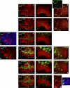
Figure 1
Figure 1
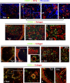
APCs expressing different markers colonize distinct areas of the human axillary lymph node. Frozen lymph node sections were probed with antibodies detecting CD3 and CD21 to identify the T lymphocyte–rich paracortex and follicles, respectively, thus providing anatomical definition (A,B). Within the paracortex, T lymphocytes were densely packed in some regions and more diffuse in others (A,B). Immunohistochemistry illustrated that cells expressing CD1a were either concentrated in diffuse T-lymphocyte zones surrounding the dense T-lymphocyte regions (C) or evenly distributed throughout areas densely packed with T lymphocytes (D). Cells expressing low and high levels of CD1a were detected (D, inset). CD208+ cells colonized similar areas to cells expressing CD1a (E,F). Dense bodies of cells expressing CD68, CD209, CD206, and CD14 were located in the medullary cords (G-L). Less frequent cells expressing CD68, CD206, and CD209 were also detected in the diffuse T-lymphocyte regions surrounding the medullary cords and follicles (G-K). Cells expressing CD68 or CD14 were also detected in the follicles (G,L). Rare BDCA-2+ plasmacytoid APCs were detected in the paracortex (M). Panels A to C, E, G, H, J, and L were acquired from the same area on sequential sections as were panels D, F, I, and K. Blue represents DAPI staining of cell nuclei (A,M, inset). *Background autofluorescence. Data are representative of 5 independent experiments. BDCA-2+ APC data are representative of 3 independent experiments.
Figure 2
Figure 2
APC populations expressing a similar phenotype to cutaneous migratory APCs were detected in the paracortex of the human axillary lymph node. Frozen lymph node sections were probed with antibodies detecting CD3 and CD21 to identify the T lymphocyte–rich paracortex and follicles, respectively (A). Immunohistochemistry illustrated that a subset of CD1a+ APCs in the paracortex (B) coexpressed CD207 or Langarin (C). CD207+ APCs were either concentrated in the diffuse T-lymphocyte zones surrounding dense T-lymphocyte regions (B,C) or distributed throughout areas densely packed with T lymphocytes (D). Proportions of both CD1a+CD207 and CD1a+CD207+ APCs coexpressed CD208/DC-LAMP; however, not all CD208+ cells expressed CD1a or CD207 (E). APCs expressing CD1a or CD208 did not coexpress CD68 (F). Differing expression patterns of CD1a, CD207, and CD208 can therefore be used to distinguish between 3 APC populations in the paracortex: CD1a+CD207CD208+/−, CD1a+CD207+CD208+/−, and CD208+CD1aCD207 APCs (G). Subsets of CD1a+CD207 and CD1a+CD207+ APCs (H) and the majority of CD1aCD208+ APCs (I) expressed the lipid presentation molecule CD1b. Cells expressing CD1b but no CD208 or CD1a were rare (I). Panels A to C, E, F, and I were acquired from the same area on sequential sections. The images shown in panel H were acquired from different fields. *Background autofluorescence. Data are representative of 5 independent experiments.
Figure 3
Figure 3
APCs expressing a similar phenotype to the APCs in medullary cords lined the capsule and trabeculae in the human axillary lymph node. Immunohistochemistry demonstrated that the APCs packed into the medullary cords expressed CD209; the majority of these APCs also expressed CD206, CD14, and CD68 (A-C). Anatomical definition was obtained by staining lymph node sections with antibodies detecting CD3 and CD21 to identify the T lymphocyte–rich paracortex and follicles, respectively (D). A sequential section probed with antibodies detecting CD209 and CD14 demonstrated that a population expressing these markers was scattered along the basement membranes of the capsule and trabeculae (E,F). This subcapsular APC population was consistently observed in all axillary lymph nodes (H) and coexpressed CD68 (G,I); a few of these cells also expressed CD206 (J). In one axillary lymph node, an unusually large number of cells expressing CD209 were detected beneath the capsule and along the trabeculae (K). Panels A to C were acquired from the same area on sequential sections as were panels D to F. M indicates medullary cords; white arrows identify the capsule (C) and trabeculae (T) (D-G, K). *Background autofluorescence. Data are representative of 4 independent experiments.
Figure 4
Figure 4
APCs expressing a similar phenotype to the APCs in medullary cords were also detected in the paracortex of the human axillary lymph node. Anatomical definition was obtained by staining frozen lymph node sections with antibodies detecting CD3 and CD21 to identify the T lymphocyte–rich paracortex and follicles, respectively (A). Sequential sections were probed with a panel of antibodies to defining APC markers (B-G). In addition to the CD209+ APCs in the medullary cords, cells expressing low-level CD209 colonized the diffuse T-lymphocyte areas surrounding the medullary cords and follicles (B-G). The majority of CD209lo APCs in the diffuse T-lymphocyte zones coexpressed CD68 (B), whereas CD14 (C) and CD206 (D) expression was variable. These CD209lo APCs did not express CD1a (E), CD207 (F), CD208 (G), or BDCA-2 (H). BDCA-2+ APCs also lacked CD68 expression (I,J). Panels A to G were acquired from the same area on sequential sections. Blue represents DAPI staining of cell nuclei (A,H-J). Data are representative of 4 independent experiments. BDCA-2+ APC data are representative of 3 independent experiments.
Figure 5
Figure 5
Conduits branch from the capsule and trabeculae and radiate into the human axillary lymph node. Immunohistochemistry illustrated that collagen fibers expressing fibronectin branched from the outer capsule of the lymph node and integrated with the fibroreticular cell network in the lymph node (A). Similarly, trabeculae that were constructed of collagen fibers and expressed fibronectin radiated deep into the lymph node, branched, and merged with the conduit network (B). Data are representative of 4 independent experiments.
Figure 6
Figure 6
APCs in the human axillary lymph node associate with the fibroreticular cell network. Frozen lymph node sections were probed with antibodies to either collagen or fibronectin to map the fibroreticular cell network. Multicolor immunohistochemistry illustrated that the majority of CD1a+CD207, CD1a+CD207+ (A,B), and CD208+ APCs (C-E) were either in intimate contact with or tethered to fibroreticular cell structures. Most APCs expressing CD209 (F-I) and CD14 (J-L) were also in close contact with fibroreticular cell structures. Interestingly, CD209+ and CD14+ APCs tended to cluster around ring-like structures resembling high endothelial venules (F-J). Just beneath the outer capsule, there was a narrow void lacking collagen fibers; the CD209+ and CD14+ APCs distributed along the basement membrane of the capsule were tethered to the collagen fibers at either side of this subcapsular space, possibly sinus (F,G,K,L). Data are representative of 3 independent experiments.

References

    1. Villadangos JA, Schnorrer P. Intrinsic and cooperative antigen-presenting functions of dendritic-cell subsets in vivo. Nat Rev Immunol. 2007;7:543–555. - PubMed
    1. Allan RS, Waithman J, Bedoui S, et al. Migratory dendritic cells transfer antigen to a lymph node-resident dendritic cell population for efficient CTL priming. Immunity. 2006;25:153–162. - PubMed
    1. Henri S, Vremec D, Kamath A, et al. The dendritic cell populations of mouse lymph nodes. J Immunol. 2001;167:741–748. - PubMed
    1. Kissenpfennig A, Henri S, Dubois B, et al. Dynamics and function of Langerhans cells in vivo: dermal dendritic cells colonize lymph node areas distinct from slower migrating Langerhans cells. Immunity. 2005;22:643–654. - PubMed
    1. Allan RS, Smith CM, Belz GT, et al. Epidermal viral immunity induced by CD8alpha+ dendritic cells but not by Langerhans cells. Science. 2003;301:1925–1928. - PubMed

Publication types