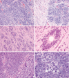
Embryonal tumors with abundant neuropil and true rosettes: a distinctive CNS primitive neuroectodermal tumor
- PMID: 18987548
- PMCID: PMC4512670
- DOI: 10.1097/PAS.0b013e318186235b
Embryonal tumors with abundant neuropil and true rosettes: a distinctive CNS primitive neuroectodermal tumor
Abstract
Embryonal neoplasms of the central nervous system (CNS) generally arise in the early years of life and behave in a clinically aggressive manner, but vary somewhat in their microscopic appearance. Several groups have reported examples of an embryonal tumor with combined histologic features of ependymoblastoma and neuroblastoma, a lesion referred to as "embryonal tumor with abundant neuropil and true rosettes" (ETANTR). Herein, we present 22 new cases, and additional clinical follow-up on our 7 initially reported cases, to better define the histologic features and clinical behavior of this distinctive neoplasm. It affects infants and arises most often in cerebral cortex, the cerebellum and brainstem being less frequent sites. Unlike other embryonal tumors of the CNS, girls are more commonly affected than boys. On neuroimaging, the tumors appear as large, demarcated, solid masses featuring patchy or no contrast enhancement. Five of our cases (18%) were at least partly cystic. Distinctive microscopic features include a prominent background of mature neuropil punctuated by true rosettes formed of pseudo-stratified embryonal cells circumferentially disposed about a central lumen (true rosettes). Of the 25 cases with available follow-up, 19 patients have died, their median survival being 9 months. Performed on 2 cases, cytogenetic analysis revealed extra copies of chromosome 2 in both. We believe that the ETANTR represents a histologically distinctive form of CNS embryonal tumor.
Figures


References
-
- Dunham C, Sugo E, Tobias V, et al. Embryonal tumor with abundant neuropil and true rosettes (ETANTR): report of a case with prominent neurocytic differentiation. J Neurooncol. 2007;84:91–98. - PubMed
-
- Eberhart CG. In search of the medulloblast: neural stem cells and embryonal brain tumors. Neurosurg Clin N Am. 2007;18:59–69. - PubMed
-
- Eberhart CG, Brat DJ, Cohen KJ, et al. Pediatric neuroblastic brain tumors containing abundant neuropil and true rosettes. Pediatr Dev Pathol. 2000;4:346–352. - PubMed
-
- Fuller C, Fouladi M, Gajjar A, et al. Chromosome 17 abnormalities in pediatric neuroblastic tumor with abundant neuropil and true rosettes. Am J Clin Pathol. 2006;126:277–283. - PubMed
-
- Giangaspero F, Eberhart CG, Haapasalo H, et al. Medulloblastoma. In: Louis DN, Ohgaki H, Wiestler OD, et al., editors. WHO Classification of Tumors of the Central Nervous System. IARC Press; Lyon: 2007. pp. 132–140.
MeSH terms
Grants and funding
LinkOut - more resources
Full Text Sources
Medical

