. 2008 Dec 18;456(7224):985-8.
doi: 10.1038/nature07444. Epub 2008 Nov 5.

X-ray structure of NS1 from a highly pathogenic H5N1 influenza virus

Affiliations

X-ray structure of NS1 from a highly pathogenic H5N1 influenza virus

Zachary A Bornholdt et al. Nature. .

Abstract

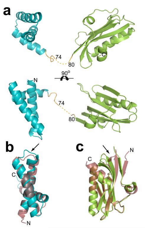
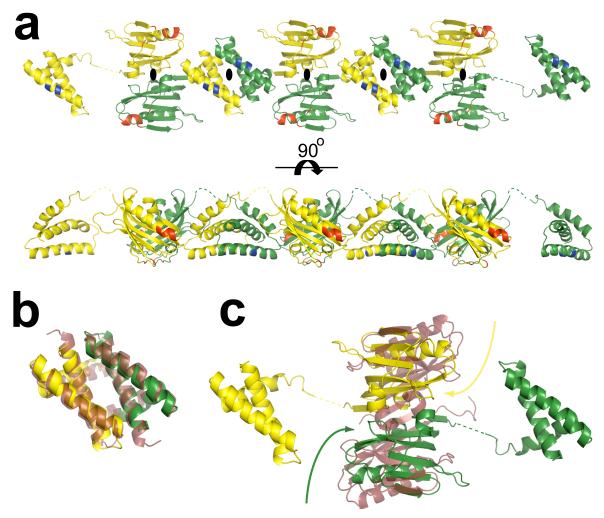
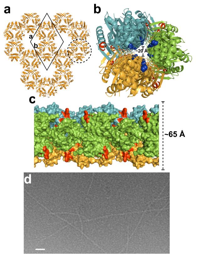
The recent emergence of highly pathogenic avian (H5N1) influenza viruses, their epizootic and panzootic nature, and their association with lethal human infections have raised significant global health concerns. Several studies have underlined the importance of non-structural protein NS1 in the increased pathogenicity and virulence of these strains. NS1, which consists of two domains-a double-stranded RNA (dsRNA) binding domain and the effector domain, separated through a linker-is an antagonist of antiviral type-I interferon response in the host. Here we report the X-ray structure of the full-length NS1 from an H5N1 strain (A/Vietnam/1203/2004) that was associated with 60% of human deaths in an outbreak in Vietnam. Compared to the individually determined structures of the RNA binding domain and the effector domain from non-H5N1 strains, the RNA binding domain within H5N1 NS1 exhibits modest structural changes, while the H5N1 effector domain shows significant alteration, particularly in the dimeric interface. Although both domains in the full-length NS1 individually participate in dimeric interactions, an unexpected finding is that these interactions result in the formation of a chain of NS1 molecules instead of distinct dimeric units. Three such chains in the crystal interact with one another extensively to form a tubular organization of similar dimensions to that observed in the cryo-electron microscopy images of NS1 in the presence of dsRNA. The tubular oligomeric organization of NS1, in which residues implicated in dsRNA binding face a 20-A-wide central tunnel, provides a plausible mechanism for how NS1 sequesters varying lengths of dsRNA, to counter cellular antiviral dsRNA response pathways, while simultaneously interacting with other cellular ligands during an infection.


Figures

Figure 1
Figure 1. H5N1 NS1 structure
a. Cartoon representation of the H5N1 NS1 structure as viewed looking across (top) and down onto (bottom) the main α-helix of the RBD (aquamarine) that is implicated in dsRNA binding. The ED is colored in green, and the linker region in orange along with the 5 residues (75-79) not well defined in the electron density map shown in dotted line. b. Structural alignment of H5N1 NS1 RBD (aquamarine) with H3N1 NS1 RBD (ruby, pdb id: 1AIL), and (c) alignment of the H5N1 NS1 ED (green) with H1N1 NS1 ED (ruby, pdb id: 2GX9). In both b and c the alignments are oriented to display the areas of greatest deviation, the V22F conformational change in a loop region observed in the H5N1 RBD in panel b, and the movement of the β-sheets in the H5N1 ED in panel c, are indicated by the black arrows.
Figure 2
Figure 2. RBD and ED dimer formation and NS1 chain
a. The RBD and ED of each NS1 molecule separately interact with their respective domains from the neighboring NS1 molecules, related by crystallographic 2-fold axes (perpendicular to plane of the paper indicated the black ovals), resulting in the formation of a chain of NS1 with alternating RBD and ED dimers. The 2-fold related NS1 molecules are colored separately in yellow and green. The residues critical to dsRNA (residues 38, and 41) and CPSF (conserved residues F103, M106, and GLEWN183-187) binding,, are colored in blue and red respectively. b. Superposition of H5N1 RBD dimer with the H3N2 RBD dimer (in ruby, pdb id: 1AIL), each protomeric subunit in the H5N1 RBD dimer is colored differently in yellow and green. c. Structural alignment of the H5N1 dimer and H1N1 NS1 ED dimer (in ruby, pdb id: 2GX9) demonstrating the twisting motion (curved arrows) of the H5N1 ED monomers, with respect to H1N1 ED, toward their RBDs. Each monomer in the H5N1 NS1 dimer is colored as in panel a. The dimeric interface of the H5N1 NS1 ED, with a buried surface area of ~1600 Å2, consists of a series of electrostatic interactions: a salt bridge between K131 and E97, hydrogen bonding involving the side chains of T91 and R193, E196 and R200, E152 and the amide group of L95, as well as a back bone hydrogen bond between the E96 amide group and the E152 carbonyl group. In contrast, the dimeric interactions in the H1N1 NS1 ED consists of primarily strong hydrophobic interactions along the continuous anti-parallel β-sheet involving residues L90, V136, and L141.
Figure 3
Figure 3. NS1 tubular structure
a. A thin slice of the crystal packing of H5N1 NS1 molecules (orange) as viewed along the crystallographic c-axis. The crystallographic unit cell (solid black diamond) along with the a and b axes are shown for reference. One of the tubular structures formed by the interaction of three NS1 chains related by the crystallographic 31-screw axis (perpendicular to the plane of the paper) is shown surrounded by a broken circle. b. NS1 tubular structure (diameter of ~65 Å) as viewed down its long axis showing the ~20 Å wide tunnel. Each of the three chains is colored differently in green, orange and light blue. The residues 38 and 41 critical for dsRNA binding are shown as blue spheres, demonstrating their location within the central tunnel, the residues critical to CPSF binding,, are colored red. c. A surface representation of the tubular structure (diameter ~65 Å) viewed across its long axis. The CSPF binding sites colored in red are surface exposed. Each chain is colored as in panel b. d. A cryo-electron micrograph of native H1N1 NS1 in the presence of dsRNA. Long tubular structures of diameter ~70 Å can be clearly visualized. Scale bar, ~500 Å.

References

    1. Abdel-Ghafar AN, et al. Update on avian influenza A (H5N1) virus infection in humans. N Engl J Med. 2008;358:261–273. - PubMed
    1. Beigel JH, et al. Avian influenza A (H5N1) infection in humans. N Engl J Med. 2005;353:1374–1385. - PubMed
    1. Seo SH, Hoffmann E, Webster RG. The NS1 gene of H5N1 influenza viruses circumvents the host anti-viral cytokine responses. Virus Res. 2004;103:107–113. - PubMed
    1. Seo SH, Hoffmann E, Webster RG. Lethal H5N1 influenza viruses escape host anti-viral cytokine responses. Nat Med. 2002;8:950–954. - PubMed
    1. Chien CY, et al. A novel RNA-binding motif in influenza A virus non-structural protein 1. Nat Struct Biol. 1997;4:891–895. - PubMed

Publication types

MeSH terms

Substances

Associated data