

Recurrent translocations involving the IRF4 oncogene locus in peripheral T-cell lymphomas
- PMID: 18987657
- PMCID: PMC2656414
- DOI: 10.1038/leu.2008.320
Recurrent translocations involving the IRF4 oncogene locus in peripheral T-cell lymphomas
Abstract
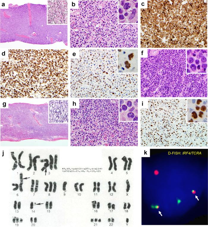
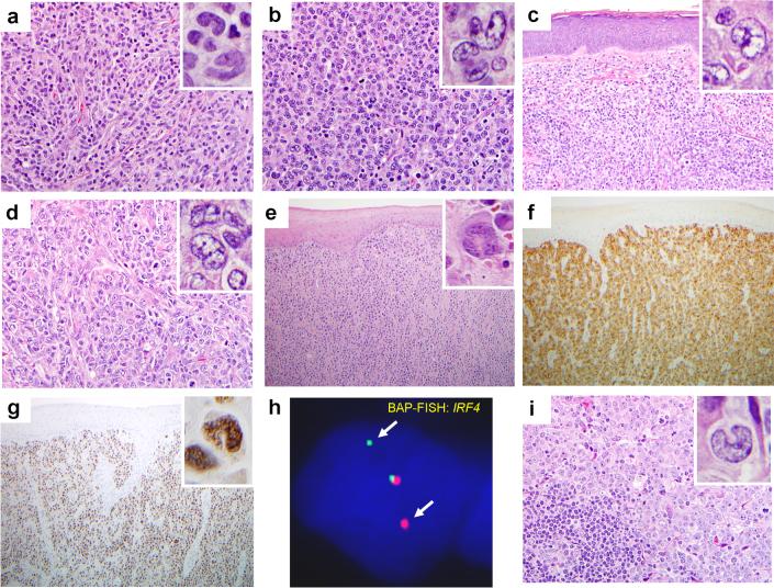
Oncogenes involved in recurrent chromosomal translocations serve as diagnostic markers and therapeutic targets in hematopoietic tumors. In contrast to myeloid and B-cell neoplasms, translocations in peripheral T-cell lymphomas (PTCLs) are poorly understood. Here, we identified recurrent translocations involving the multiple myeloma oncogene-1/interferon regulatory factor-4 (IRF4) locus in PTCLs. IRF4 translocations exist in myeloma and some B-cell lymphomas, but have not been reported earlier in PTCLs. We studied 169 PTCLs using fluorescence in situ hybridization and identified 12 cases with IRF4 translocations. Two cases with t(6;14)(p25;q11.2) had translocations between IRF4 and the T-cell receptor-alpha (TCRA) locus. Both were cytotoxic PTCLs, unspecified (PTCL-Us) involving bone marrow and skin. In total, 8 of the remaining 10 cases were cutaneous anaplastic large-cell lymphomas (ALCLs) without TCRA rearrangements (57% of cutaneous ALCLs tested). These findings identified IRF4 translocations as a novel recurrent genetic abnormality in PTCLs. Cytotoxic PTCL-Us involving bone marrow and skin and containing IRF4/TCRA translocations might represent a distinct clinicopathologic entity. Translocations involving IRF4 but not TCRA appear to occur predominantly in cutaneous ALCLs. Detecting these translocations may be useful in lymphoma diagnosis. Further, due to its involvement in translocations, MUM1/IRF4 protein may play an important biologic role in some PTCLs, and might represent a possible therapeutic target.
Figures
References
-
- Jaffe ES, Harris NL, Stein H, Vardiman J. Pathology and Genetics of Tumours of Haematopoietic and Lymphoid Tissues. IARC Press; Lyon, France: 2001.
-
- Savage KJ, Chhanabhai M, Gascoyne RD, Connors JM. Characterization of peripheral T-cell lymphomas in a single North American institution by the WHO classification. Ann Oncol. 2004;15:1467–1475. - PubMed
-
- Streubel B, Vinatzer U, Willheim M, Raderer M, Chott A. Novel t(5;9)(q33;q22) fuses ITK to SYK in unspecified peripheral T-cell lymphoma. Leukemia. 2006;20:313–318. - PubMed
-
- Li R, Morris SW. Development of anaplastic lymphoma kinase (ALK) small-molecule inhibitors for cancer therapy. Med Res Rev. 2008;28:372–412. - PubMed
Publication types
MeSH terms
Substances
Grants and funding
LinkOut - more resources
Full Text Sources
Other Literature Sources
Medical
