. 2009 Feb;129(2):438-48.
doi: 10.1038/jid.2008.279. Epub 2008 Nov 6.

An essential role for dermal primary cilia in hair follicle morphogenesis

Affiliations

An essential role for dermal primary cilia in hair follicle morphogenesis

Jonathan M Lehman et al. J Invest Dermatol. 2009 Feb.

Abstract

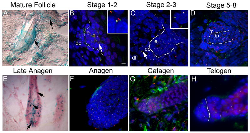
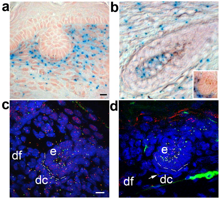
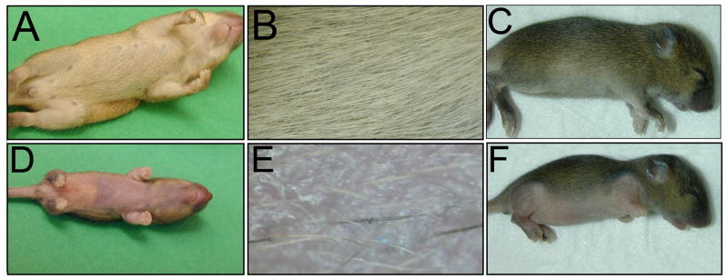
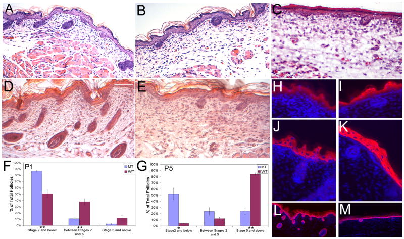
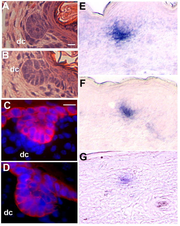
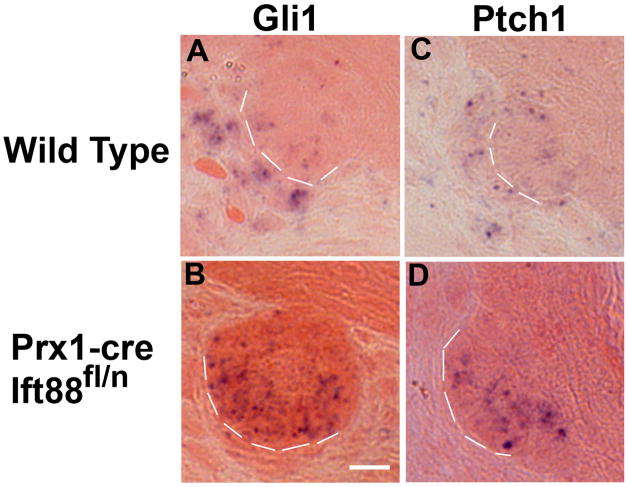
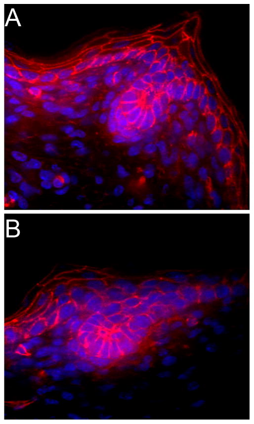
The primary cilium is a microtubule-based organelle implicated as an essential component of a number of signaling pathways. It is present on cells throughout the mammalian body; however, its functions in most tissues remain largely unknown. Herein we demonstrate that primary cilia are present on cells in murine skin and hair follicles throughout morphogenesis and during hair follicle cycling in postnatal life. Using the Cre-lox system, we disrupted cilia assembly in the ventral dermis and evaluated the effects on hair follicle development. Mice with disrupted dermal cilia have severe hypotrichosis (lack of hair) in affected areas. Histological analyses reveal that most follicles in the mutants arrest at stage 2 of hair development and have small or absent dermal condensates. This phenotype is reminiscent of that seen in the skin of mice lacking Shh or Gli2. In situ hybridization and quantitative RT-PCR analysis indicates that the hedgehog pathway is downregulated in the dermis of the cilia mutant hair follicles. Thus, these data establish cilia as a critical signaling component required for normal hair morphogenesis and suggest that this organelle is needed on cells in the dermis for reception of signals such as sonic hedgehog.


Conflict of interest statement

Conflict of Interest

The authors state no conflict of interest.

Figures

Figure 1
Figure 1
Temporal and spatial analysis of an IFT88/Tg737 reporter gene (A, E) and protein (B–D, and F–H) reveals the presence of a primary cilium on epithelial and mesenchymal cells of the skin and hair follicle in embryos and adults. (A, E) Ift88 expression was analyzed in heterozygous Ift88tm1Rpw (Ift88Δ2–3-β-Gal) mice using a β-galactosidase reporter gene by X-Gal staining of skin sections. In the mature follicle (A) and late anagen follicle (E) and in the developing skin and hair, Ift88 expression (blue) is detected in most cell types. Expression is prominent in the (arrow) cortex and (indented arrow) ORS (epithelium), (arrowhead) dermal papilla (mesenchyme) (E), and in cells near the (arrow) bulge region (stem cells) of the hair follicle as well as the (arrowhead) interfollicular epidermis (A). (B–D,F–H) IFT88 (red) localizes to primary cilia as shown by immunofluorescence co-localization obtained using antibodies against the ciliary axoneme marker, acetylated α-tubulin (green). The nuclei (blue) were labeled with Hoechst. (B) Analysis in a stage 1–2 developing hair follicle shows primary cilia on cells of both the forming dermal condensate (dc) and epidermal placode (e). The dashed line indicates the region of the basement membrane and the intersection between the epidermis and dermis. The insert shows a magnified image of the region indicated by the arrow. (C) In a stage 2–3 developing hair follicle, cilia were present on the epidermal and dermal portion of the hair germ. The epidermal (e), dermal condensate (dc) and dermal fibroblast (df) cells are indicated. The insert shows a magnification of the region corresponding to the arrow in the dermal fibroblasts. (D) Analysis in a later stage (5–8) follicle shows primary cilia present on cells of both the matrix (m) and dermal papilla (dp). (F–H) Immunofluorescence co-localization of acetylated α-tubulin (green) and IFT88 (red) during hair follicle cycling shows that a single primary cilium is present on most cells at (F) anagen, (G) most dermal cells in catagen with reduced epidermal cilia, and (H) most cells in telogen. Scalebar is 10 microns. 3-D confocal images of cilia in the developing (1) or mature anagen (2) follicle can be seen in supplemental movies 1 and 2.
Figure 2
Figure 2
Cre activity and disruption of cilia in Prx1-cre mice is restricted to the dermal compartment of the skin and hair follicles covering the limbs, flanks, and ventrum. (A) Cre activity in the skin and stage-2 hair follicle from the ventrum of a Prx1-cre;R26R mouse is present only in dermal cells, as revealed by the lacZ reporter gene (blue). (B) Cre activity is also seen in dermal components of advanced follicles including the dermal papilla of an advanced follicle and a stage 2 follicle (inset) from a P5 Prx1-cre;R26R mouse. (C) In wild-type control mice, primary cilia are present in the epidermal (e) and dermal (dc, dermal condensate; df, dermal fibroblast) compartments of the skin and stage 2 hair follicle, as shown by co-localization of antibodies raised against acetylated α-tubulin (green) and IFT88 (red). (D) In Prx1-cre;Ift88fl/n conditional mutant mice, primary cilia are ablated from most dermal condensate (dc) and dermal fibroblast (df) cells, but cilia are unaffected on epidermal (e) cells. Rare dermal cilia are unaffected, likely due to incomplete cre activity (arrow). Scalebar is 10 microns.
Figure 3
Figure 3
Conditional disruption of primary cilia in the dermis of the ventral skin in Prx1-cre;Ift88fl/n and Prx1-cre; Kif3afl/n mice results in hypotrichosis. The pelages of (A,B,C) wild-type littermate control mice, (D,E) a Prx1-cre;Ift88fl/n, and (F) Prx1-cre; Kif3afl/n conditional mutant mouse are shown at P14. (B,E) Higher magnification images of the ventrum of the mice in A and D show that the mutant skin has an extremely sparse coat and a few follicles that appear normal. The conditional cilia mutant mice also have deformed limbs that were described previously (Haycraft et al., 2007).
Figure 4
Figure 4
Hair follicle morphogenesis is arrested at stage 2 (hair germ) in mice with primary cilia ablated from dermal cells of the skin and hair follicles. Histological sections of skin from (A) P1 wildtype, (B) mutant P1 Prx1-cre;Ift88fl/n,and (C) P1 Prx1-cre; Kif3afl/n mice show fewer and less advanced follicles in the mutant mice. This phenotype worsens at P5 in (E) mutant animals when compared to age matched (D) wildtype controls. Histomorphometric analyses of the hair follicle phenotypes in skins harvested from mutant and control mice at (F) P1 and (G) P5 indicate that most follicles arrest at stage 2 of morphogenesis in Prx1-cre;Ift88fl/n mice. The analyses were performed using a minimum of 40 longitudinal follicles in each group from (F) P1 (n=5 pairs) and (G) P5 (n=3 pairs) mice. Error bars represent SEM. Statistical comparisons were conducted using the two-tailed independent Student’s t test. * p < 0.05; ** p < 0.01. (H–M) Defects in differentiation were analyzed by immunofluorescence using antibodies against (H,I) loricrin (granular layer), (J,K) K1 (stratum spinosum), and (L,M) K5 (basal layer) of the epidermis reveal no overt differences in staining between (H,J,L) WT and (I,K,M) MT interfollicular epidermis. Scale bar is 50 microns.
Figure 5
Figure 5
Disruption of cilia in the dermis of Prx1-cre;Ift88fl/n conditional mutant mice results in a marked reduction or absence of cells in the dermal condensate of stage 2 hair follicles. Histological analysis of (A and B) Hematoxylin and Eosin stained sections and (C and D) thick section immunofluorescence analysis using epidermal marker Keratin 5 (red) antibodies shows a marked reduction in the number of cells in the dermal condensate (dc) of (B and D) Prx1-cre;Ift88fl/n conditional mutants compared to (A and C) controls. The reduced number of dermal condensate cells was also evident in (F and G) Prx1-cre;Ift88fl/n conditional mutants when compared to (E) controls as determined by staining for endogenous alkaline phosphatase activity. (E, F) are stage 2 follicles from P1 mice and (G) is a stage 2 follicle at P5. Scalebar is 10 microns.
Figure 6
Figure 6
Ablation of dermal cilia in the skin of Prx1-cre;Ift88fl/n conditional mutant mice results in downregulation of the hedgehog responsive genes, Gli1 and Patched1, during hair follicle morphogenesis. Analysis of the hedgehog signaling pathway was performed in sections of P5 skin by in situ hybridization to detect expression of the hedgehog responsive genes (A, B) Gli1 and (C, D) Patched1 (Ptch1) in (A, C) control and (B, D) Prx1-cre;Ift88fln conditional mutant mice. The mutants show reduced or absent expression for both genes in the dermal condensates and an increase in the epidermal cells. Dashed lines separate the epidermal components of the hair follicle from the underlying dermal condensate. Scalebar is 10 microns.
Figure 7
Figure 7
There are no overt changes in Canonical Wnt signaling in dermal cilia conditional mutants as determined by thick section immunofluorescent confocal analysis of β-catenin (red) localization between (A) wild type and (B) Prx1-cre;Ift88fl/n mutant mice at P1. Nuclei are stained with Hoescht (blue).

Comment in

References

    1. Benzing T, Walz G. Cilium-generated signaling: a cellular GPS? Curr Opin Nephrol Hypertens. 2006;15:245–249. - PubMed
    1. Blanpain C, Fuchs E. Epidermal Stem Cells of the Skin. Annu Rev Cell Dev Biol 2005 - PMC - PubMed
    1. Bonner JC, Osornio-Vargas AR, Badgett A, Brody AR. Differential proliferation of rat lung fibroblasts induced by the platelet-derived growth factor-AA, -AB, and -BB isoforms secreted by rat alveolar macrophages. Am J Respir Cell Mol Biol. 1991;5:539–547. - PubMed
    1. Botchkarev VA, Paus R. Molecular biology of hair morphogenesis: development and cycling. J Exp Zoolog B Mol Dev Evol. 2003;298:164–180. - PubMed
    1. Cano DA, Murcia NS, Pazour GJ, Hebrok M. Orpk mouse model of polycystic kidney disease reveals essential role of primary cilia in pancreatic tissue organization. Development. 2004;131:3457–3467. - PubMed

Publication types

MeSH terms