

Histomorphometric measurements of bone turnover, mineralization, and volume
- PMID: 18988700
- PMCID: PMC3152285
- DOI: 10.2215/CJN.04301206
Histomorphometric measurements of bone turnover, mineralization, and volume
Abstract
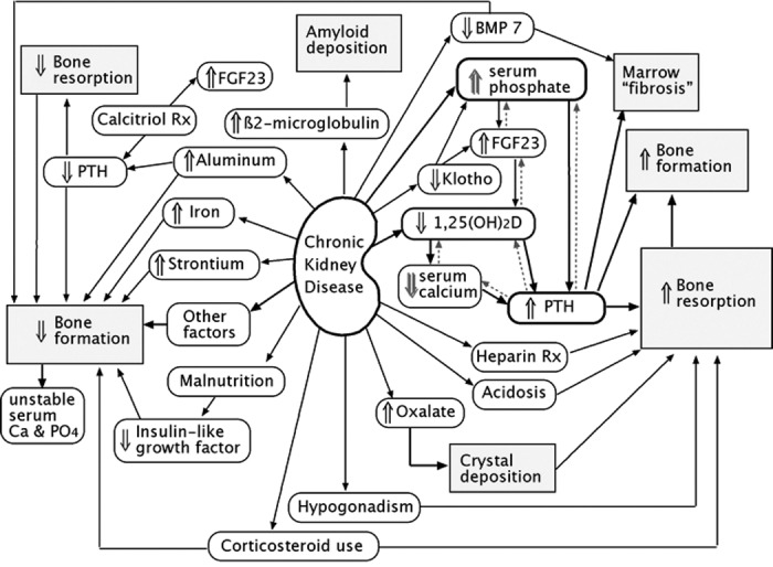
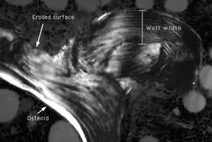
A recent Kidney Disease: Improving Global Outcomes report suggested that bone biopsies in patients with chronic kidney disease should be characterized by determining bone turnover, mineralization, and volume. This article focuses on the calculations and interpretation of these measurements. In most cases of renal osteodystrophy, the bone formation rate is roughly similar to the bone resorption rate; therefore, the bone formation indices can be used to describe turnover. It is important to remember that these conventions will not apply in some situations. Activation frequency should not be confused with bone formation rate or bone metabolic unit birth rate. Abnormal mineralization can be described using the osteoid volume, increased osteoid maturation time, or increased mineralization lag time. The concept of bone volume is the easiest to understand, but there is a large error from one biopsy to the next in the same person. There are some difficulties with each of the measurements, and further research in patients with chronic kidney must be done to enable a consensus to be reached about cut points to define categories within the spectrum of renal osteodystrophy and how to evaluate treatment responses.
Figures
References
-
- Elder G: Pathophysiology and recent advances in the management of renal osteodystrophy. J Bone Miner Res 17 :2094 –2105,2002 - PubMed
-
- Ferreira A: Development of renal bone disease. Eur J Clin Invest 36 [Suppl 2]:2 –12,2006 - PubMed
-
- Hruska KA, Teitelbaum SL: Renal osteodystrophy. N Engl J Med 333 :166 –174,1995 - PubMed
-
- Sherrard DJ, Hercz G, Pei Y, Greenwood C, Manuel A, Saiphoo C, Fenton SS, Segre GV: The spectrum of bone disease in end-stage renal failure: An evolving disorder. Kidney Int 43 :436 –442,1993 - PubMed
-
- Cunningham J, Sprague SM, Cannata-Andia J, Coco M, Cohen-Solal M, Fitzpatrick L, Goltzmann D, Lafage-Proust MH, Leonard M, Ott S, Rodriguez M, Stehman-Breen C, Stern P, Weisinger J, Osteoporosis Work Group: Osteoporosis in chronic kidney disease. Am J Kidney Dis 43 :566 –571,2004 - PubMed
Publication types
MeSH terms
LinkOut - more resources
Full Text Sources
Medical
