

Technical approach to iliac crest biopsy
- PMID: 18988702
- PMCID: PMC3152282
- DOI: 10.2215/CJN.00460107
Technical approach to iliac crest biopsy
Abstract
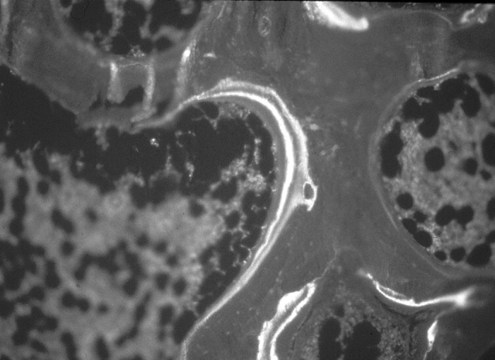
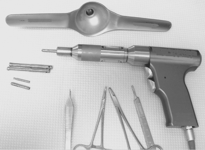
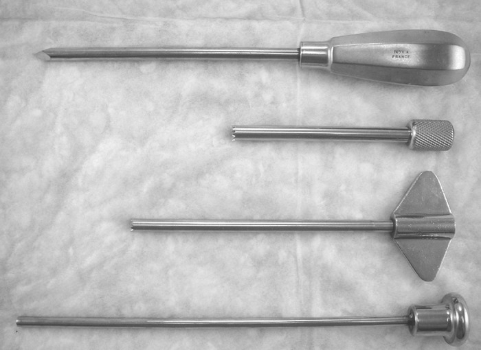
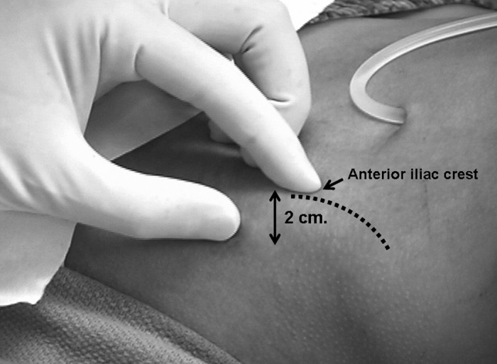
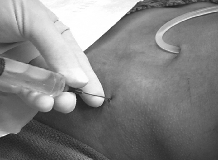
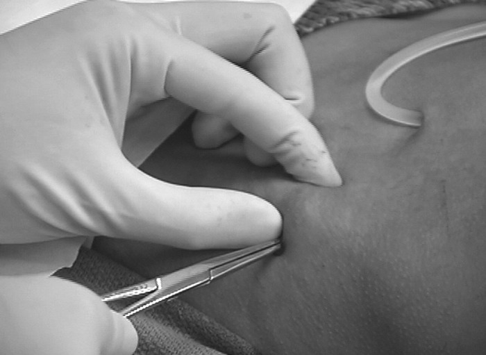
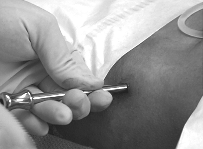
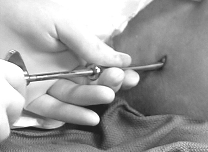
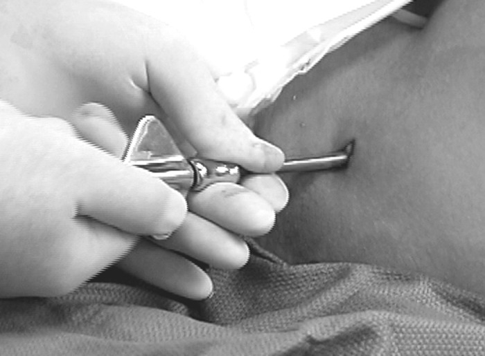
Bone histomorphometry has been the gold standard in the evaluation and diagnosis of renal osteodystrophy. The recent new definition of renal osteodystrophy as chronic kidney disease-mineral and bone disorder has once again highlighted the use of bone biopsy as a powerful and diagnostic tool to determine skeletal abnormalities in chronic kidney disease. The procedure of iliac crest bone biopsy has been proved safe and associated with very minimal morbidity. In this review, the clinical indications, preparation, instrumentation, and potential complications are discussed. Because current biochemical markers are poor predictors of bone turnover, volume, and mineralization, a wider use of bone biopsy and histomorphometry will lead to a better understanding of the bone and mineral disorders that are associated with chronic kidney disease.
Figures
References
-
- Salusky IB, Coburn JW, Brill J, Foley J, Slatopolsky E, Fine RN, Goodman WG: Bone disease in pediatric patients undergoing dialysis with CAPD or CCPD. Kidney Int 33 :975 –982,1988 - PubMed
-
- Weinstein RS: Clinical use of bone biopsy. In: Disorders of Bone and Mineral Metabolism, 2nd Ed., edited by Coe FL, Favus MJ, Philadelphia, Lippincott Williams & Wilkins,2002. , pp448 –468
-
- Monier-Faugere M-C, Langub MC, Malluche HH: Bone biopsies: A modern approach. In: Metabolic Bone Diseases and Clinically Related Disorders, 3rd Ed., edited by Avioli LV, Krane SM, San Diego, Academic Press,1998. , pp237 –273
-
- Ketteler M, Gross ML, Ritz E: Calcification and cardiovascular problems in renal failure. Kidney Int Suppl S120 –S127,2005 - PubMed
-
- Ketteler M, Westenfeld R, Schlieper G, Brandenburg V: Pathogenesis of vascular calcification in dialysis patients. Clin Exp Nephrol 9 :265 –270,2005 - PubMed
Publication types
MeSH terms
Substances
Grants and funding
LinkOut - more resources
Full Text Sources
Medical
