. 2008 Dec;173(6):1891-901.
doi: 10.2353/ajpath.2008.080378. Epub 2008 Nov 6.

Transgenic induction of vascular endothelial growth factor-C is strongly angiogenic in mouse embryos but leads to persistent lymphatic hyperplasia in adult tissues

Affiliations

Transgenic induction of vascular endothelial growth factor-C is strongly angiogenic in mouse embryos but leads to persistent lymphatic hyperplasia in adult tissues

Marja Lohela et al. Am J Pathol. 2008 Dec.

Abstract

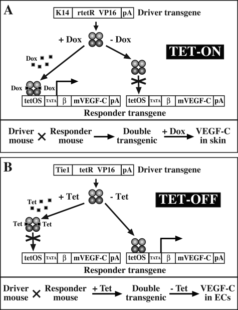
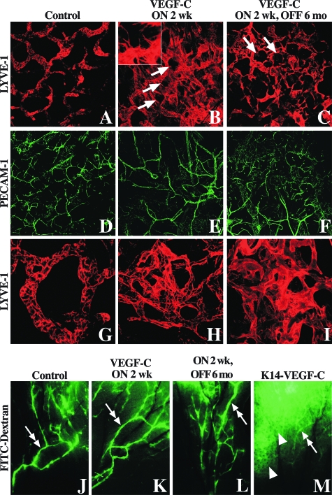
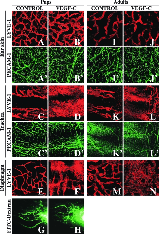
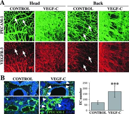
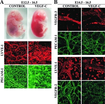
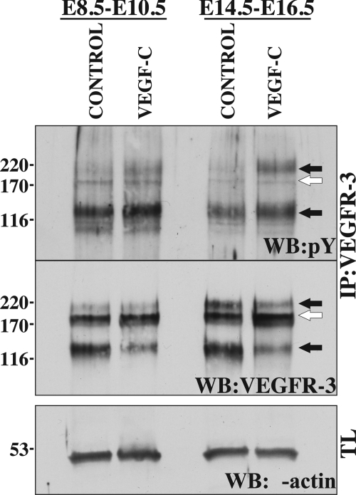
Vascular endothelial growth factor-C (VEGF-C) is the quintessential lymphangiogenic growth factor that is required for the development of the lymphatic system and is capable of stimulating lymphangiogenesis in adults by activating its receptor, VEGFR-3. Although VEGF-C is a major candidate molecule for the development of prolymphangiogenic therapy for defective lymphatic vessels in lymphedema, the stability of lymph vessels generated by exogenous VEGF-C administration is not currently known. We studied VEGF-C-stimulated lymphangiogenesis in inducible transgenic mouse models in which growth factor expression can be spatially and temporally controlled without side effects, such as inflammation. VEGF-C induction in adult mouse skin for 1 to 2 weeks caused robust lymphatic hyperplasia that persisted for at least 6 months. VEGF-C induced lymphangiogenesis in numerous tissues and organs when expressed in the vascular endothelium in either neonates or adult mice. Very few or no effects were observed in either blood vessels or collecting lymph vessels. Additionally, VEGF-C stimulated lymphangiogenesis in embryos after the onset of lymphatic vessel development. Strikingly, a strong angiogenic effect was observed after VEGF-C induction in vascular endothelium at any point before embryonic day 16.5. Our results indicate that blood vessels can undergo VEGF-C-induced angiogenesis even after down-regulation of VEGFR-3 in embryos; however, transient VEGF-C expression in adults can induce long-lasting lymphatic hyperplasia with no obvious side effects on the blood vasculature.


Figures

Figure 1
Figure 1
Schematic representation of the transgenic Tet-on and Tet-off VEGF-C expression models. For inducible VEGF-C expression, mice carrying the tissue-specific driver and VEGF-C responder transgenes are mated to obtain double-transgenic offspring. A: The K14-rtTA driver construct produces a transcriptional activator fusion protein that binds to the Tet operator sequence in the responder construct only in the presence of tetracycline antibiotics. In double-transgenic K14-rtTA/TET-VEGF-C mice, the VEGF-C responder construct is silent, and expression can be activated by administration of tetracycline or doxycycline. B: Tie1-tTA and VEC-tTA driver constructs produce a transcriptional activator fusion protein that binds to the Tet operator sequence in the responder construct only in the absence of tetracycline antibiotics. In double-transgenic Tie1-tTA/TET-VEGF-C or VEC-tTA/TET-VEGF-C mice, the VEGF-C responder construct is constitutively active. VEGF-C expression can be suppressed by administration of tetracycline or doxycycline, and induced again by withdrawal of the antibiotic.
Figure 2
Figure 2
Hyperplastic lymph vasculature induced by VEGF-C is maintained after removal of stimulation. Double-transgenic K14-rtTA/TET-VEGF-C mice were given doxycycline for 2 weeks, after which some mice were sacrificed and analyzed (B, E, H, K), and some were maintained without doxycycline for 6 months (C, F, I, L) before analysis of ear skin by fluorescent dextran lymphangiography and whole-mount immunostaining of lymphatic (LYVE-1, red) and blood vessels (PECAM-1, green). Compared to double-transgenic control littermates that did not receive doxycycline (A and G), 2-week stimulation resulted in robust lymphangiogenesis, lymphatic sprouting, and hyperplasia (B and H, inset in B shows lymphatic sprouts at higher magnification). Arrows point to the locations of some hair follicles, around which the hyperplastic plexus has formed. C and I: Six months after transgene activation was stopped, the lymphatic vasculature was still very hyperplastic. The collecting lymph vessels (double arrows) appeared to function normally, as demonstrated by fluorescent dextran lymphangiography (K and L compared to J). In the constitutive transgenic K14-VEGF-C mice (M), the dextran spread into the hyperplastic lymph capillary plexus (arrowhead) before it was taken up by the collecting vessels (double arrow). No blood vessel hyperplasia was observed in any of these mice (D–F). Original magnifications: ×100 (A–F); ×400 (G–I); ×10 (J–M); ×200 (B, inset).
Figure 3
Figure 3
VEGF-C expressed in the endothelium induces lymphangiogenesis but no angiogenesis. Whole-mount staining of lymph (LYVE-1, red) and blood vessels (PECAM-1, green) in ear skin, trachea, and diaphragm of double-transgenic Tie1-tTA/TET-VEGF-C and littermate control pups (A–F, A’–D’) and adult mice (I–N, I’–L’) after VEGF-C induction; fluorescent dextran lymphangiography in the ear of Tie1-tTA/TET-VEGF-C and littermate control pups after VEGF-C induction (G, H). A–H and A’–D’: Pregnant females were maintained on doxycycline until pups were born, at which point doxycycline was removed. VEGF-C expression was induced by maintaining double-transgenic Tie1-tTA/TET-VEGF-C pups without doxycycline, and Tie1-tTA/TET-VEGF-C pups and littermate controls were sacrificed when ∼4 weeks old. Function of the collecting lymph vessels was analyzed by FITC-dextran lymphangiography. I–N and I’–L’: Pregnant females were maintained on doxycycline until pups were born, and pups were maintained on doxycycline until they reached adulthood. VEGF-C expression was induced in adult double-transgenic Tie1-tTA/TET-VEGF-C mice by removing doxycycline, and mice were then maintained without doxycycline for 2 months and analyzed together with littermate controls. Original magnifications: ×10 (G, H); ×100 (A–F, I–N, A’–D’, I’–L’).
Figure 4
Figure 4
Endothelial VEGF-C induces embryonic angiogenesis. Whole-mount staining of blood vessels (A) and quantification of endothelial cells in cryosections (B) from E10.5 Tie1-tTA/TET-VEGF-C and littermate control embryos after VEGF-C induction. A: Pregnant females were maintained on tetracycline until approximately E6.5, at which point tetracycline was removed to induce VEGF-C expression in double-transgenic embryos. Tie1-tTA/TET-VEGF-C and control embryos were analyzed at approximately E10.5 by whole-mount staining for PECAM-1 (green) and VEGFR-3 (red). Arrows point to large vessels in control embryos in which VEGFR-3 expression has been down-regulated. B: A pregnant female was maintained on tetracycline until approximately E7.5, at which point tetracycline was removed to induce VEGF-C expression in double-transgenic embryos, and embryos were collected at approximately E10.5. Cryosections of Tie1-tTA/TET-VEGF-C and control embryos were stained for the blood vascular marker PECAM-1 and 4,6-diamidino-2-phenylindole for nuclei. PECAM-1-positive endothelial cells were counted from equivalent vessel cross-sections from the jugular area in Tie1-tTA/TET-VEGF-C and control embryos. Arrowheads point to regions with vessel hyperplasia. Error bars represent standard deviations between the average endothelial cell numbers in individual embryos; ***P < 0.001. Original magnifications: ×100 (A; B, top); ×400 (B, bottom).
Figure 5
Figure 5
Endothelial VEGF-C affects both lymph and blood vessels in late embryogenesis. Representative photographs and whole-mount staining of vascular markers from skins of E16.5 Tie1-tTA/TET-VEGF-C and control embryos after VEGF-C induction. A: Pregnant females were maintained on tetracycline until approximately E12.5, at which point tetracycline was removed to induce VEGF-C expression in double-transgenic embryos. Photos were taken and skins from Tie1-tTA/TET-VEGF-C and control embryos were analyzed at approximately E16.5 by whole-mount stainings of lymphatic (LYVE-1, red) and blood vessels (PECAM-1, green). B: Pregnant females were taken off tetracycline at E14.5 to induce VEGF-C expression in double-transgenic embryos, and skins from Tie1-tTA/TET-VEGF-C and control embryos were analyzed at approximately E16.5 by whole-mount stainings of VEGFR-3 (red) and PECAM-1 (green) or LYVE-1 (red) and VEGFR-2 (green). Original magnifications: ×100 (A); ×400 (B).
Figure 6
Figure 6
Endothelial VEGF-C overexpression induces tyrosine phosphorylation, synthesis, and degradation of VEGFR-3 in embryos. Analysis of VEGFR-3 tyrosine phosphorylation in Tie1-tTA/TET-VEGF-C and control embryos after VEGF-C induction. Pregnant females were maintained on tetracycline until it was removed to induce VEGF-C expression in double-transgenic embryos at approximately E8.5 or E14.5. Tie1-tTA/TET-VEGF-C and control embryos were harvested after 2 days of induction. VEGFR-3 was immunoprecipitated from pooled lysates of whole embryos at E10.5 and of the lungs of E16.5 embryos. The immunocomplexes were separated on SDS-PAGE, Western blotted, and probed with anti-phosphotyrosine antibody (top). The blots were then stripped and reprobed with anti-VEGFR-3 antibodies (middle). Aliquots from the same lysates were separated on SDS-PAGE, Western blotted, and probed for β-actin to confirm equal amounts of protein in the lysates (bottom). Arrows in the top and middle panels point to the 195-kDa, 175-kDa (open arrow), and 125-kDa forms of VEGFR-3. Note the decreased amount but increased relative phosphorylation of the 125-kDa VEGFR-3 receptor band. IP, immunoprecipitation; WB, Western blot; TL, total lysate.

Similar articles

Cited by

References

    1. Carmeliet P, Ferreira V, Breier G, Pollefeyt S, Kieckens L, Gertsenstein M, Fahrig M, Vandenhoeck A, Harpal K, Ebenhardt C, Declercq C, Pawling J, Moons L, Collen D, Risau W, Nagy A. Abnormal blood vessel development and lethality in embryos lacking a single VEGF allele. Nature. 1996;380:435–439. - PubMed
    1. Ferrara N, Carver-Moore K, Chen H, Dowd M, Lu L, O’Shea KS, Powell-Braxton L, Hillan KJ, Moore MW. Heterozygous embryonic lethality induced by targeted inactivation of the VEGF gene. Nature. 1996;380:439–442. - PubMed
    1. Karkkainen MJ, Haiko P, Sainio K, Partanen J, Taipale J, Petrova TV, Jeltsch M, Jackson DG, Talikka M, Rauvala H, Betsholtz C, Alitalo K. Vascular endothelial growth factor C is required for sprouting of the first lymphatic vessels from embryonic veins. Nat Immunol. 2004;5:74–80. - PubMed
    1. Benest AV, Harper SJ, Herttuala SY, Alitalo K, Bates DO. VEGF-C induced angiogenesis preferentially occurs at a distance from lymphangiogenesis. Cardiovasc Res. 2008;78:315–323. - PMC - PubMed
    1. Oh SJ, Jeltsch MM, Birkenhager R, McCarthy JE, Weich HA, Christ B, Alitalo K, Wilting J. VEGF and VEGF-C: specific induction of angiogenesis and lymphangiogenesis in the differentiated avian chorioallantoic membrane. Dev Biol. 1997;188:96–109. - PubMed

Publication types

MeSH terms

Substances