. 2008 Nov 7;322(5903):953-6.
doi: 10.1126/science.1164840.

Insights into translational termination from the structure of RF2 bound to the ribosome

Affiliations

Insights into translational termination from the structure of RF2 bound to the ribosome

Albert Weixlbaumer et al. Science. .

Abstract

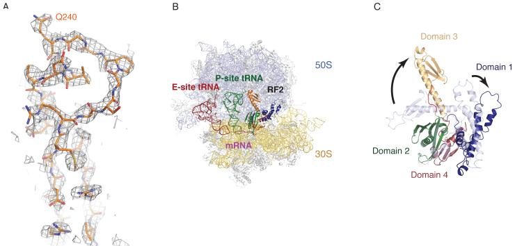
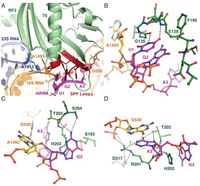
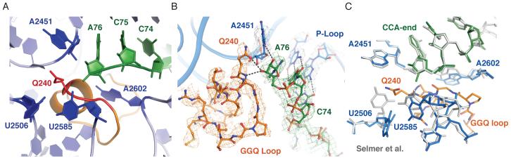
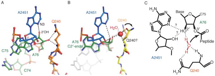
The termination of protein synthesis occurs through the specific recognition of a stop codon in the A site of the ribosome by a release factor (RF), which then catalyzes the hydrolysis of the nascent protein chain from the P-site transfer RNA. Here we present, at a resolution of 3.5 angstroms, the crystal structure of RF2 in complex with its cognate UGA stop codon in the 70S ribosome. The structure provides insight into how RF2 specifically recognizes the stop codon; it also suggests a model for the role of a universally conserved GGQ motif in the catalysis of peptide release.


Figures

Fig. 1
Fig. 1
The structure of release factor RF2 in the ribosome. A. Unbiased difference Fourier maps showing density for RF2 in the peptidyl transferase center of the ribosome. B. Overview of the structure showing the 50S subunit (light blue), the 30S subunit (yellow) with E-site tRNA (red), P-site tRNA (green) and RF2 colored by domains as in C. The mRNA is shown in magenta. C. Conformational differences between the isolated crystal structure of RF2 (15) (shown in light blue), and the ribosome bound form (colored by domains as labeled) are indicated. Loop regions connecting domain 3 with domain 2 and 4, that undergo substantial conformational changes, are highlighted in red.
Fig. 2
Fig. 2
Interaction of RF2 with the stop codon in the decoding center. A. Overview of the decoding center showing the UGA stop codon (magenta). Domain 2 of RF2 is shown in green, except for parts interacting with the codon, which are shown in red. Key bases from 16S RNA and A1913 of 23S RNA are also shown. (B to D). Details of interactions at the first (B), second (C) and third (D) positions of the stop codon with elements of RF2 and the decoding center.
Fig 3
Fig 3
Interaction of RF2 with the peptidyl transferase center. A. Overview of the peptidyl transferase center showing the terminal CCA of P-site tRNA (green), the conserved GGQ motif of RF2 (red) and key bases of 23S RNA (blue). B. Interaction of the GGQ loop of RF2 (orange) with the terminal ribose of P-site tRNA (green), showing sigma A weighted 3mFobs – 2DFcalc maps, where m is the figure of merit, D is the sigma A weight, and Fobs and Fcalc are the observed and calculated structure factors, respectively. Potential hydrogen bonds between the conserved Q240 and the ribose of P-site tRNA are shown. C. Changes in the peptidyl transferase center upon RF2 binding compared to the 70S structure with an empty A site (gray) (12). The structure shows that RF2 would clash with elements of 23S RNA and induces conformational changes, in particular in U2506 and U2585.
Fig. 4
Fig. 4
A model of the substrate complex suggesting the basis for catalysis. A. The interaction of Q240 of RF2 and A2451 of 23S RNA in the current structure. B. A proposed structure showing how a minor change in the orientation of Q240 would allow it to coordinate a water molecule (transparent orange versus yellow). In gray is a transition-state analog superposed on this structure [1VQ7, taken from (24)] that was used to place the putative water that would take part in nucleophilic attack on the ester bond. C. A schematic representation of how a network of interactions among the conserved Q240, the coordinated water molecule that is the attacking nucleophile, the ribose of A76 of P-site tRNA (in C2′-endo conformation) and A2451 would act to facilitate catalysis.

Comment in

References

    1. Capecchi MR. Proc Natl Acad Sci U S A. 1967;58:1144. - PMC - PubMed
    1. Youngman EM, McDonald ME, Green R. Annu Rev Microbiol. 2008;62:353. - PubMed
    1. Single-letter abbreviations for the amino acid residues are as follows: A, Ala; C, Cys; D, Asp; E, Glu; F, Phe; G, Gly; H, His; I, Ile; K, Lys; L, Leu; M, Met; N, Asn; P, Pro; Q, Gln; R, Arg; S, Ser; T, Thr; V, Val; W, Trp; and Y, Tyr.

    1. Frolova LY, et al. RNA. 1999;5:1014. - PMC - PubMed
    1. Ito K, Uno M, Nakamura Y. Nature. 2000;403:680. - PubMed

Publication types

MeSH terms