Review
. 2008 Nov 15;112(10):3939-48.
doi: 10.1182/blood-2008-07-161166.

Red cell membrane: past, present, and future

Affiliations
Review

Red cell membrane: past, present, and future

Narla Mohandas et al. Blood. .

Abstract

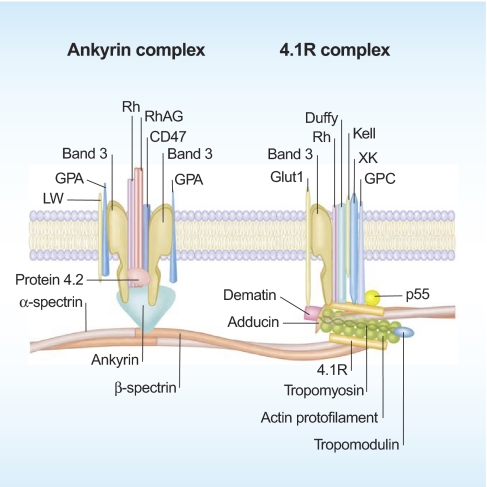
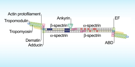
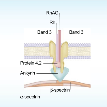
As a result of natural selection driven by severe forms of malaria, 1 in 6 humans in the world, more than 1 billion people, are affected by red cell abnormalities, making them the most common of the inherited disorders. The non-nucleated red cell is unique among human cell type in that the plasma membrane, its only structural component, accounts for all of its diverse antigenic, transport, and mechanical characteristics. Our current concept of the red cell membrane envisions it as a composite structure in which a membrane envelope composed of cholesterol and phospholipids is secured to an elastic network of skeletal proteins via transmembrane proteins. Structural and functional characterization of the many constituents of the red cell membrane, in conjunction with biophysical and physiologic studies, has led to detailed description of the way in which the remarkable mechanical properties and other important characteristics of the red cells arise, and of the manner in which they fail in disease states. Current studies in this very active and exciting field are continuing to produce new and unexpected revelations on the function of the red cell membrane and thus of the cell in health and disease, and shed new light on membrane function in other diverse cell types.


Figures

None
Narla Mohandas
None
Patrick G. Gallagher
Figure 1
Figure 1
Multilobular reticulocyte (top left panel), the precursor of the mature discoid red cell (top right panel). A red cell traversing from the splenic cord to splenic sinus (bottom left panel). Note the marked deformation the cell undergoes during its passage through the narrow endothelial slit separating the cord from the sinus. Ellipsoidal cells generated in vitro by flow-induced deformation in vitro of discoid cells (bottom right panel).
Figure 2
Figure 2
A schematic representation of red cell membrane. The membrane is a composite structure in which a plasma membrane envelope composed of amphiphilic lipid molecules is anchored to a 2-dimensional elastic network of skeletal proteins through tethering sites (transmembrane proteins) embedded in the lipid bilayer. Illustration by Paulette Dennis.
Figure 3
Figure 3
The horizontal linkages between spectrin-spectrin dimers and between spectrin, actin, and protein 4.1R in the junctional complex in the spectrin-based membrane skeleton. The repeats of α-spectrin are colored gray while those of β-spectrin are colored light green. The single helical repeat at N-terminus of α-spectrin of 1 dimer interacts with the 2 helical repeat at C-terminus of β-spectrin of the second dimer to constitute spectrin dimer-dimer interaction indicated in pink. The PS binding spectrin repeats are colored in dark blue while repeats with low thermal stability (Tm < 37°C) are shown in red. The various components of the junctional complex at the end of spectrin dimer are also shown. EF hands at C-terminus of α-spectrin and the actin-binding domain (ABD) at N-terminus of β-spectrin are also indicated. Illustration by Paulette Dennis.
Figure 4
Figure 4
Red cell morphology. Hereditary spherocytosis (HS; top panel); nonhemolytic hereditary elliptocytosis (HE; middle panel); elliptocytes, poikilocytes, and fragmented red cells in hemolytic HE (bottom panel).
Figure 5
Figure 5
Membrane defects in HS affect the “vertical” interactions anchoring the membrane skeleton to the lipid bilayer. Deficiency in any one of the protein components (band 3, RhAG, ankyrin, protein 4.2, or spectrin) involved in the anchoring process leads to HS. Illustration by Paulette Dennis.

References

    1. Van Leeuwenhoek A. Other microscopical observations made by the same, about the texture of the blood, the sap of some plants, the figures of sugar and salt, and the probable cause of the difference of their tastes. Philos Trans R Soc Lond. 1675;10:380–385.
    1. Bessis M, Delpech G. Discovery of the red blood cell with notes on priorities and credits of discoveries, past, present and future. Blood Cells. 1981;7:447–480. - PubMed
    1. Gulliver G. Medical Times and Gazette. London: John Churchill & Sons; 1862. Blood of Vertebrata.
    1. Gorter E, Grendel F. On the bimolecular layers of lipids on the chromocytes of the blood. J Exp Med. 1925;41:439–443. - PMC - PubMed
    1. Singer SJ, Nicolson GL. The fluid mosaic model of the structure of cell membranes. Science. 1972;175:720–731. - PubMed

MeSH terms