. 2008 Nov;4(11):e1000197.
doi: 10.1371/journal.ppat.1000197. Epub 2008 Nov 7.

Broadening of neutralization activity to directly block a dominant antibody-driven SARS-coronavirus evolution pathway

Affiliations

Broadening of neutralization activity to directly block a dominant antibody-driven SARS-coronavirus evolution pathway

Jianhua Sui et al. PLoS Pathog. 2008 Nov.

Abstract

Phylogenetic analyses have provided strong evidence that amino acid changes in spike (S) protein of animal and human SARS coronaviruses (SARS-CoVs) during and between two zoonotic transfers (2002/03 and 2003/04) are the result of positive selection. While several studies support that some amino acid changes between animal and human viruses are the result of inter-species adaptation, the role of neutralizing antibodies (nAbs) in driving SARS-CoV evolution, particularly during intra-species transmission, is unknown. A detailed examination of SARS-CoV infected animal and human convalescent sera could provide evidence of nAb pressure which, if found, may lead to strategies to effectively block virus evolution pathways by broadening the activity of nAbs. Here we show, by focusing on a dominant neutralization epitope, that contemporaneous- and cross-strain nAb responses against SARS-CoV spike protein exist during natural infection. In vitro immune pressure on this epitope using 2002/03 strain-specific nAb 80R recapitulated a dominant escape mutation that was present in all 2003/04 animal and human viruses. Strategies to block this nAb escape/naturally occurring evolution pathway by generating broad nAbs (BnAbs) with activity against 80R escape mutants and both 2002/03 and 2003/04 strains were explored. Structure-based amino acid changes in an activation-induced cytidine deaminase (AID) "hot spot" in a light chain CDR (complementarity determining region) alone, introduced through shuffling of naturally occurring non-immune human VL chain repertoire or by targeted mutagenesis, were successful in generating these BnAbs. These results demonstrate that nAb-mediated immune pressure is likely a driving force for positive selection during intra-species transmission of SARS-CoV. Somatic hypermutation (SHM) of a single VL CDR can markedly broaden the activity of a strain-specific nAb. The strategies investigated in this study, in particular the use of structural information in combination of chain-shuffling as well as hot-spot CDR mutagenesis, can be exploited to broaden neutralization activity, to improve anti-viral nAb therapies, and directly manipulate virus evolution.


Conflict of interest statement

The authors have declared that no competing interests exist.

Figures
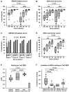
Figure 1
Figure 1. Serologic analysis.
A, 11 of convalescent serum samples were obtained from SARS patients of the 2002/03 outbreak in China. Neutralizing activities of these serum samples were analyzed against pseudotyped viruses bearing the S protein of Tor2 or GD03 at indicated dilutions. Data were shown in a box and whiskers graph. The box extends from 25th percentile to the 75th percentile, with a line at the median. The whiskers above and below the box indicate the 95th and 5th percentiles. The dots above and below the box showed highest and lowest data points. A symbol of “*” indicates the data points of a SARS-CoV negative healthy human serum sample. IC90 for each patient serum was calculated and the statistic analysis was done with IC90 value using one way ANOVA for correlated samples. The same data presentation and the statistic analysis methods were used for the following panels B and D. B, Neutralization assay of Tor2 and GD03 pseudotyped viruses with six civet serum samples collected from animal market in Guangzhou in Jan. 2004. Similarly as Fig. 1A, “*” indicates the data points for a SARS-CoV negative civet cat serum sample. C, Neutralization assay of Tor2 and GD03 pseudotyped viruses with four serum samples from 2003/04 outbreak. The samples labeled in the graph as patient 1–4 that were the same patients described before . D. Ten serum samples from civet cat farmers collected in June 2003in Guangdong Province were analyzed against Tor2 and GD03 pseudotyped viruses. E, Tor2 -RBD binding activity of 2002/03, 2003/04 outbreak, and Civet cat farmers' serum samples were analyzed by ELISA. These serum samples were the same as those used in Panel A, C, and D, respectively. Dilution of serum was 1∶180. The data was presented the same way as panel A and unpaired Student t-test was used for statistical analysis. The average and standard deviation (SD) of background binding of two SARS-CoV negative human serum samples (Ctrl.HS) were also shown. F, A comparison of the competition ability of different serum samples for 80R's binding to Tor2-RBD were evaluated by ELISA. Dilution for all samples was 1∶20. The non-specific competition for 80R's binding to Tor-RBD by two Ctrl. HS or one control civet cat serum (Ctrl.CS) was also shown. Serum samples were corresponding to those used in panel A, B, C or D. The data presentation and statistic analysis were done the same way as panel E.
Figure 2
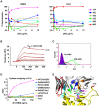
Figure 2. Neutralization of pseudotyped viral infection by anti-GD03 Abs and epitope mapping of GD03 nAb 11A.
A, Five anti-GD03 Abs isolated by phage Ab library screening against purified GD03-RBD were analyzed for neutralizing activity using GD03 (Left panel) or Tor2 (Right panel) spike pseudoyped viruses. B, Kinetic characterization of GD03-RBD binding to 11A-IgG1. Abs were captured on a CM4 chip via immobilized anti-human IgG1 on the chip. GD03-RBD at various concentrations (2 folds serial dilutions, highest concentration was indicated) was injected over the chip surface. Binding kinetics was evaluated using a 1∶1 interaction model. In each panel, the binding response curves (red lines) are overlaid with the fit of the interaction model (black lines). All ka, kd, KD value showed in the table represent the means and standard errors of three experiments. C, Competition of 11A for the binding of GD03-RBD-C9 to 293T-ACE2 cells. GD03-RBD-C9 or control-RBD-C9 (Filled purple) used for staining is at 20 ug/mL and the Abs (256 or 80R negative control) were used at 50 ug/mL to compete for the binding of GD03-RBD to 293T-ACE2 cells. D, Epitope mapping of 11A. Purified proteins of a set of GD03-RBD or Tor2-RBD mutants were coated to ELISA plates at indicated concentrations. 2 µg/ml of 11A-IgG1 followed by HRP-anti-human IgG1 were used to detect the binding of 11A with different mutants. E, Interface of structure of the Tor2- RBD/80R complex 21. S1-RBD is in yellow. CDR loops (H1-H3 and L1-L3) of 80R, amino acids of 80R-CDRL1 and L2 (162–164 and 182) as well as 472L and 480D of S1-RBD are colored as shown.
Figure 3
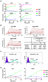
Figure 3. Characterization of Ab 256.
A, Neutralization of pseudoviral infection by 256-IgG1. An anti-CXCR4 Ab 33-IgG1 was used as a negative control, 80R and 11A were used as positive control for Tor2- and GD03-viruses, respectively. B, Kinetic characterization of the binding of spike RBDs to 256-IgG1. Binding kinetics was evaluated similarly as described in Fig. 2. B. C, Competition of 256 for the binding of Tor2- or GD03-RBD-C9 to 293T-ACE2 cells. Left, competition for the binding of Tor2-RBD-Ig to 293T-ACE2 cells. 0.5 ug/mL of Tor2-RBD-Ig or control-Ig (Filled purple) used for the staining of 293T-ACE2 cells and the scFvs (control or 256) were used at 5 ug/mL to compete for the binding. Right, competition for the binding of GD03-RBD to 293T-ACE2 cells. The assay was the same as Fig. 2C except 256 was used here. D, Ab 256 Competition ELISA Assay. A fixed amount of 256 scFv expressing phages (256-phages) were mixed with various scFv-Fc antibody or full-length IgG1s at indicated antibody concentration, and the mixtures were then added to Tor2-RBD (left) or GD03-RBD (right) coated ELISA plate. The competition of 256-IgG1s for the binding of 256-phages to RBDs were determined by measuring the remaining binding of 256-phages using HRP-anti-M13 antibody. 256-phages homologous to 256-IgG1 were used as positive controls and both showed competition for 256-phage binding to Tor2-RBD and GD03-RBD, 80R or 11A did not show inhibition of 256-phages binding to either Tor2- or GD03-RBDs.
Figure 4
Figure 4. Sequence alignment of the Vk of five Abs identified from 80R-Vk-cs library and 80R.
The best-matched germline Vκ and Jκ genes of 80R are showed on top of the alignment. CDR regions are labeled in large boxes, amino acid substitutions from germline are colored in blue. A dash indicates no amino acid at that position. Amino acids 161–164 in CDRL1 were highlighted in pink. All 5 Abs have one consensus change from S to N at position 163. WRCY hot spots of AID are colored in green.
Figure 5
Figure 5. Broadly neutralizing activity of cs- and fm-Abs.
A, sequence comparison of eight Abs identified from 80R-cs or -fm library selection. *Amino acid sequence in CDRL1 (161–164). ** 80R was identified from Tor2-S1 targeted library selection ; five fm-Abs were from both D480A or D480G and GD03 targeted library selection. B, Neutralization activity of the eight Abs (full-length IgG1) was tested with the same pseudotyped viruses as used in Fig. 3A at indicated Ab concentration. Cs5, cs84, fm6 and fm39 appeared to be the four BnAbs.

References

    1. Ksiazek TG, Erdman D, Goldsmith CS, Zaki SR, Peret T, et al. A novel coronavirus associated with severe acute respiratory syndrome. N Engl J Med. 2003;348:1953–1966. - PubMed
    1. Marra MA, Jones SJ, Astell CR, Holt RA, Brooks-Wilson A, et al. The Genome sequence of the SARS-associated coronavirus. Science. 2003;300:1399–1404. - PubMed
    1. Rota PA, Oberste MS, Monroe SS, Nix WA, Campagnoli R, et al. Characterization of a Novel Coronavirus Associated with Severe Acute Respiratory Syndrome. Science. 2003;300:1394–1399. - PubMed
    1. Drosten C, Gunther S, Preiser W, van der Werf S, Brodt H-R, et al. Identification of a Novel Coronavirus in Patients with Severe Acute Respiratory Syndrome. N Engl J Med. 2003;348:1967–1976. - PubMed
    1. Liang G, Chen Q, Xu J, Liu Y, Lim W, et al. Laboratory diagnosis of four recent sporadic cases of community-acquired SARS, Guangdong Province, China. Emerg Infect Dis. 2004;10:1774–1781. - PMC - PubMed

Publication types

MeSH terms