

Cellular FLIP can substitute for the herpes simplex virus type 1 latency-associated transcript gene to support a wild-type virus reactivation phenotype in mice
- PMID: 18989818
- PMCID: PMC2980827
- DOI: 10.1080/13550280802216510
Cellular FLIP can substitute for the herpes simplex virus type 1 latency-associated transcript gene to support a wild-type virus reactivation phenotype in mice
Abstract
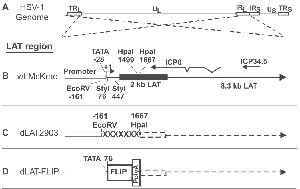
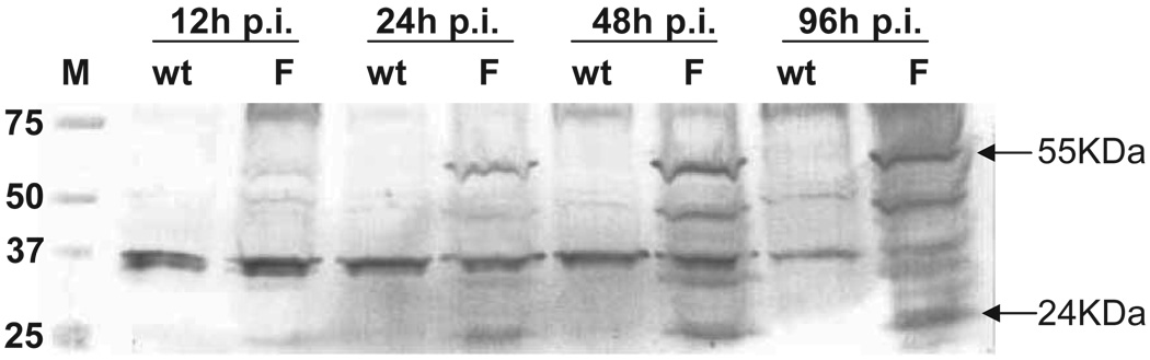
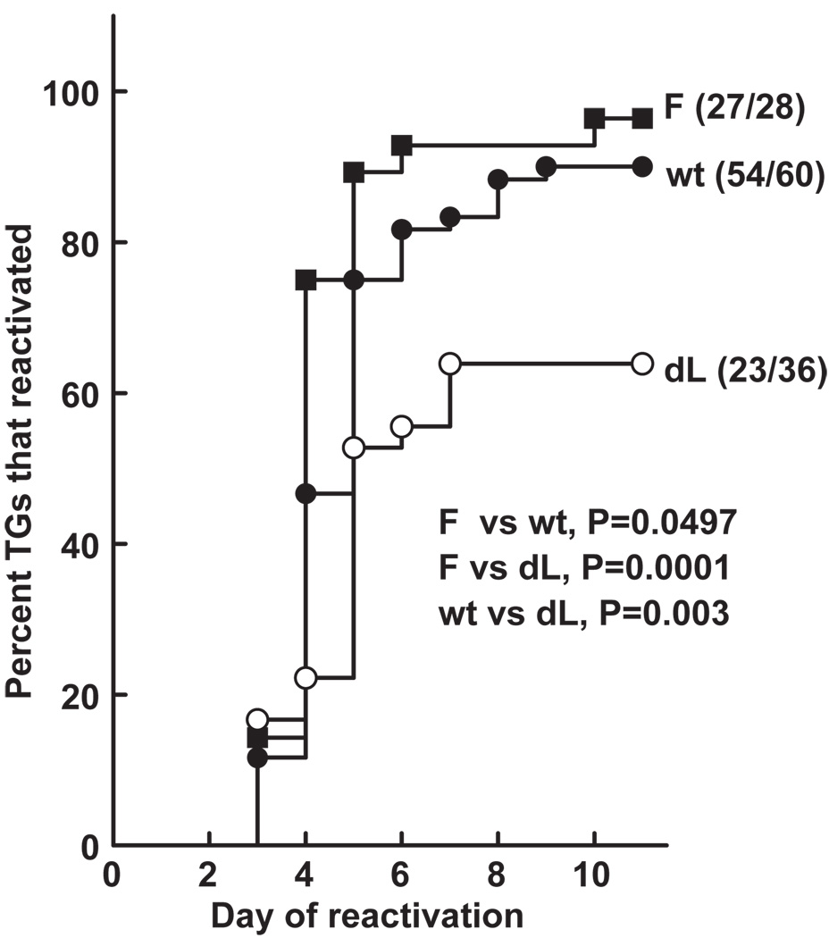
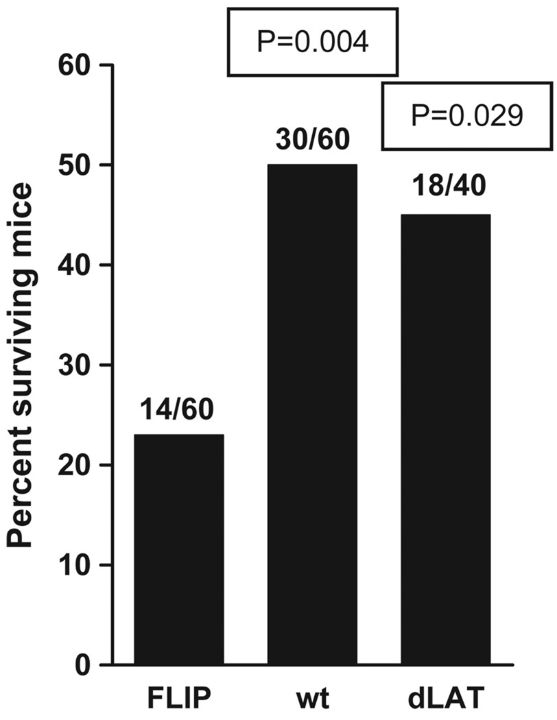
Latency-associated transcript (LAT) deletion mutants of herpes simplex virus type 1 (HSV-1) have reduced reactivation phenotypes. Thus, LAT plays an essential role in the latency-reactivation cycle of HSV-1. We have shown that LAT has antiapoptosis activity and demonstrated that the chimeric virus, dLAT-cpIAP, resulting from replacing LAT with the baculovirus antiapoptosis gene cpIAP, has a wild-type HSV-1 reactivation phenotype in mice and rabbits. Thus, LAT can be replaced by an alternative antiapoptosis gene, confirming that LAT's antiapoptosis activity plays an important role in the mechanism by which LAT enhances the virus' reactivation phenotype. However, because cpIAP interferes with both of the major apoptosis pathways, these studies did not address whether LAT's proreactivation phenotype function was due to blocking the extrinsic (Fas-ligand-, caspase-8-, or caspase-10-dependent pathway) or the intrinsic (mitochondria-, caspase-9-dependent pathway) pathway, or whether both pathways must be blocked. Here we constructed an HSV-1 LAT(-) mutant that expresses cellular FLIP (cellular FLICE-like inhibitory protein) under control of the LAT promoter and in place of LAT nucleotides 76 to 1667. Mice were ocularly infected with this mutant, designated dLAT-FLIP, and the reactivation phenotype was determined using the trigeminal ganglia explant model. dLAT-FLIP had a reactivation phenotype similar to wild-type virus and significantly higher than the LAT(-) mutant dLAT2903. Thus, the LAT function responsible for enhancing the reactivation phenotype could be replaced with an antiapoptosis gene that primarily blocks the extrinsic signaling apoptosis pathway.
Conflict of interest statement
Figures
Similar articles
-
A herpes simplex virus type 1 mutant expressing a baculovirus inhibitor of apoptosis gene in place of latency-associated transcript has a wild-type reactivation phenotype in the mouse.J Virol. 2005 Oct;79(19):12286-95. doi: 10.1128/JVI.79.19.12286-12295.2005. J Virol. 2005. PMID: 16160155 Free PMC article.
-
Reactivation phenotype in rabbits of a herpes simplex virus type 1 mutant containing an unrelated antiapoptosis gene in place of latency-associated transcript.J Neurovirol. 2007;13(1):78-84. doi: 10.1080/13550280601164333. J Neurovirol. 2007. PMID: 17454452
-
A gene capable of blocking apoptosis can substitute for the herpes simplex virus type 1 latency-associated transcript gene and restore wild-type reactivation levels.J Virol. 2002 Feb;76(3):1224-35. doi: 10.1128/jvi.76.3.1224-1235.2002. J Virol. 2002. PMID: 11773398 Free PMC article.
-
Mapping herpes simplex virus type 1 latency-associated transcript sequences that protect from apoptosis mediated by a plasmid expressing caspase-8.J Neurovirol. 2004 Aug;10(4):260-5. doi: 10.1080/13550280490468690. J Neurovirol. 2004. PMID: 15371157 Review.
-
HSV LAT and neuronal survival.Int Rev Immunol. 2004 Jan-Apr;23(1-2):187-98. doi: 10.1080/08830180490265592. Int Rev Immunol. 2004. PMID: 14690860 Review.
Cited by
-
Identification of herpes simplex virus type 1 proteins encoded within the first 1.5 kb of the latency-associated transcript.J Neurovirol. 2009 Sep;15(5-6):439-48. doi: 10.3109/13550280903296353. J Neurovirol. 2009. PMID: 20175695
-
A Journey through the Minefield of the Discovery and Characterization of Latency-Related RNA/Latency-Associated Transcript.Viruses. 2024 Sep 30;16(10):1562. doi: 10.3390/v16101562. Viruses. 2024. PMID: 39459896 Free PMC article. Review.
-
Regulation of neurotropic herpesvirus productive infection and latency-reactivation cycle by glucocorticoid receptor and stress-induced transcription factors.Vitam Horm. 2021;117:101-132. doi: 10.1016/bs.vh.2021.06.005. Epub 2021 Jul 21. Vitam Horm. 2021. PMID: 34420577 Free PMC article.
-
Small non-coding RNAs encoded within the herpes simplex virus type 1 latency associated transcript (LAT) cooperate with the retinoic acid inducible gene I (RIG-I) to induce beta-interferon promoter activity and promote cell survival.Virus Res. 2013 Aug;175(2):101-9. doi: 10.1016/j.virusres.2013.04.005. Epub 2013 May 3. Virus Res. 2013. PMID: 23648811 Free PMC article.
-
Herpes simplex virus type 1 (HSV-1)-induced apoptosis in human dendritic cells as a result of downregulation of cellular FLICE-inhibitory protein and reduced expression of HSV-1 antiapoptotic latency-associated transcript sequences.J Virol. 2010 Jan;84(2):1034-46. doi: 10.1128/JVI.01409-09. Epub 2009 Nov 11. J Virol. 2010. PMID: 19906927 Free PMC article.
References
-
- Boldin MP, Goncharov TM, Goltsev YV, Wallach D. Involvement of MACH, a novel MORT1/FADD-interacting protease, in Fas/APO-1- and TNF receptor-induced cell death. Cell. 1996;85:803–815. - PubMed
-
- Brugha R, Keersmaekers K, Renton A, Meheus A. Genital herpes infection: a review. Int J Epidemiol. 1997;26:698–709. - PubMed
Publication types
MeSH terms
Substances
Grants and funding
LinkOut - more resources
Full Text Sources
Medical
Research Materials
Miscellaneous
